

Wide field imaging energy dispersive X-ray absorption spectroscopy
Posted on January 09, 2020
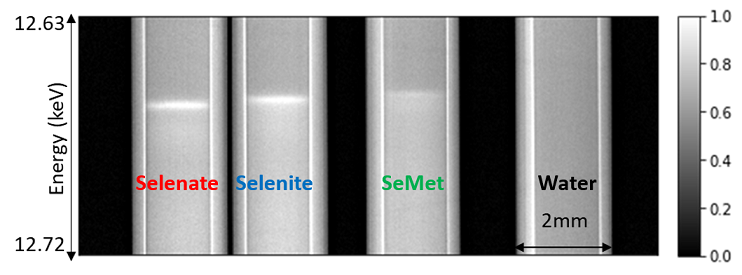
Based on spectral K-edge subtraction imaging (developed at CLS-BMIT), a new energy dispersive X-ray absorption spectroscopy method is developed at BMIT-BM beamline for simultaneous wide-field imaging and transmission X-ray Absorption Spectroscopy (XAS). Sufficient energy and spatial resolution are demonstrated for both full field imaging and computed tomography in quantifying selenium chemical species.
Read more (peng-qi-fall-2019.pdf)
3D Imaging of Hydrogen Induced Cracking in Pipeline Steel
Posted on October 07, 2019
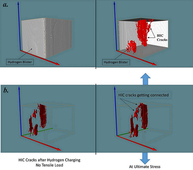
Environmental conditions of service affect the resistance of pipeline steels to failure, as a result, the applied stress which would be otherwise considered safe can be responsible for fracture due to hydrogen induced cracking (HIC). Synchrotron 3D computed tomography (CT) at BMIT-ID beamline at Canadian Light Source was used to explain the features of the HIC cracks formation during tensile tests.
Read more (mostafijur,_szpunar-summer-2019.pdf)
Visualization of 3D Bioprinted Tissue Scaffolds Using Synchrotron-Based Phase Contrast Imaging
Posted on October 07, 2019
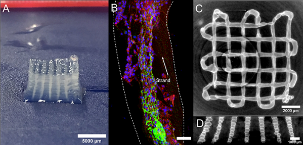
We developed the synchrotron-based x-ray inline phase contrast imaging-computed tomography (SR-inline-PCI-CT) at the Biomedical Imaging and Therapy (BMIT) Facility of Canadian Light Source to visualize the printed hydrogel-based scaffolds.
Read more (liqun_ning,_daniel_chen-summer-2019.pdf)
Optimizing Middle-Ear Prostheses
Posted on September 12, 2019
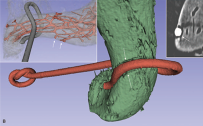
The middle ear efficiently transmits vibrations of the eardrum caused by sound entering the ear canal to fluid in the cochlea, the end organ of hearing Pathologies of the middle ear bones, the malleus, incus and stapes, can disrupt the transmission of sound energy and lead to so called conductive hearing loss.
Read more (ladak_clsi_aug_2019_feature.pdf)
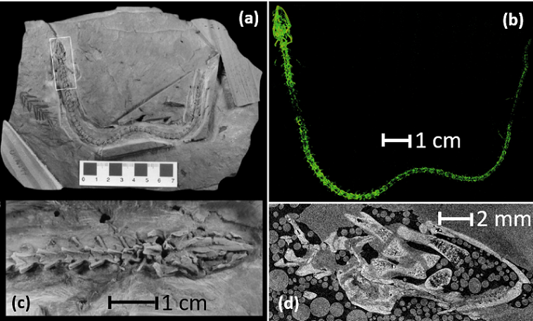
Synchrotron X-ray Micro-CT of a 60 million years old salamander
Posted on August 20, 2019
A 60 million-year-old salamander fossil was imaged (inside rock) at BMIT ID beamline at 80keV X-ray energy.
Read more (mauricio_barbi,_jim_gardner.pdf)
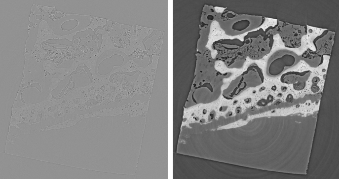
Investigation of growth at sutural sites in the presence of an external oscillatory mechanical stimulus using a swine model
Posted on April 22, 2019
Cranial sutures are the soft connective tissue that join skull bones. It is understood that a heightened stress/strain state at the suture site induces a biological response through growth and remodeling. Investigators Owen Addison and Dan Romanyk are currently working to develop a fundamental understanding of how cranial sutures respond to an increased stress/strain state.
Read more (bmit-pic-of-the-month-romanyk-28-03-2019.pdf)
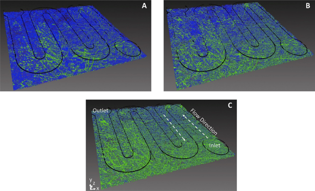
Transient, spatially resolved desaturation of gas diffusion layers measured via synchrotron visualization
Posted on April 22, 2019
The transient 3-D visualization of the desaturation process of flooded gas diffusion layers (GDLs) is presented for the first time.
Read more (bmit-pic-of-the-month-anderson-28-03-2019.pdf)
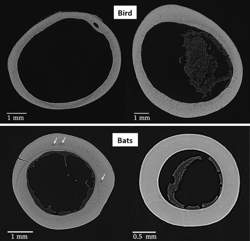
Three‐Dimensional Orientation of Vascular Canals and Cross‐Sectional Geometry of Cortical Bone in Birds and Bats
Posted on April 22, 2019
In this study researchers examined the vascular canal network in the humerus and femur of a sample of 31 bird and 24 bat species using synchrotron micro‐computed tomography (micro‐CT) to look for a connection between canal orientation and functional loading.
Read more (bmit-pic-of-the-month-pratt-28-03-2019.pdf)
Micro-Angiography in a Rat Model of Osteoarthritis
Posted on January 31, 2019
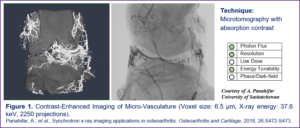
These images represent ex vivo micro-angiography, after infusion of barium/gelatine mixture, in an osteoarthritic rat 6 weeks post-surgery. Vasculature down to two voxel size in diameter can be observed.
Read more (bmit_highlights_jan2019.pdf)
Imaging the Effects of Prolonged Unloading on Osteocyte Lacunar Density in a Rat Model
Posted on January 31, 2019
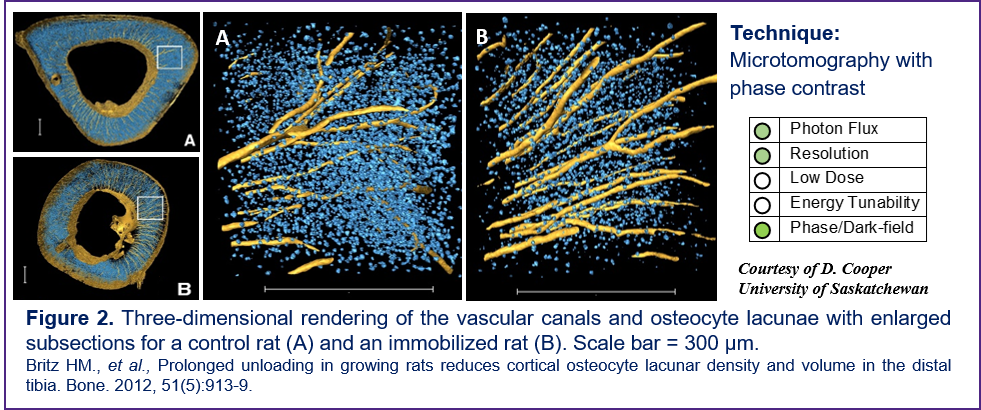
The primary objective of this study was to test the hypothesis that unloading (sciatic neurectomy) during growth results in altered osteocyte lacunar density in the tibial diaphysis of the rat. Secondarily, a potential effect of unloading on mean lacunar volume was explored.
Read more (bmit_highlights_jan2019.pdf)
In vivo Imaging of Mucus Formation in a Piglet Model of Cystic Fibrosis
Posted on January 31, 2019
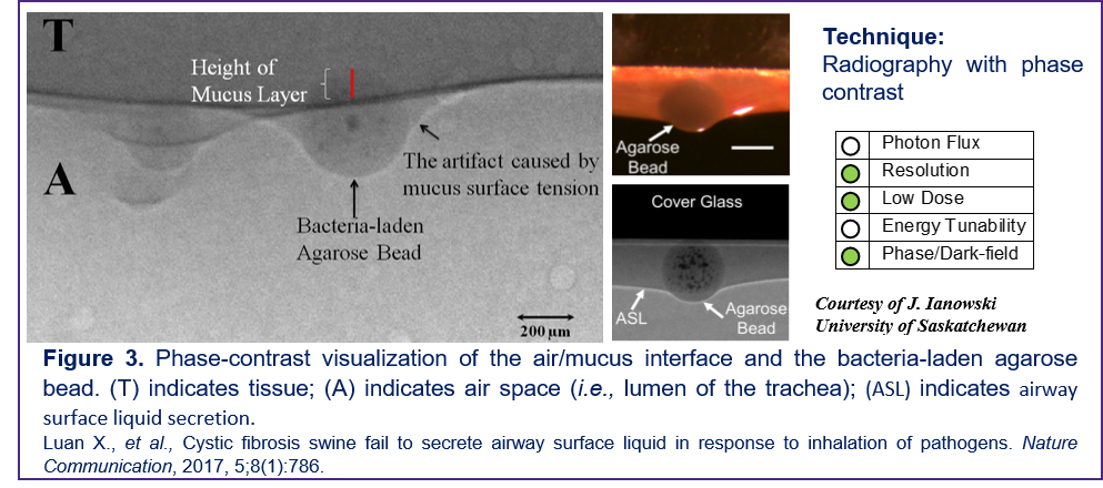
In this study the response of airway submucosal glands to inhaled bacteria and its connection to cystic fibrosis was investigated. Prior to using BMIT anatomical visualization of airway and mucus had been limited due to limitations of contrast and resolution available using conventional imaging techniques.
Read more (bmit_highlights_jan2019.pdf)
Mapping of Strontium Distribution in a Rat Model of Osteoporosis
Posted on January 31, 2019
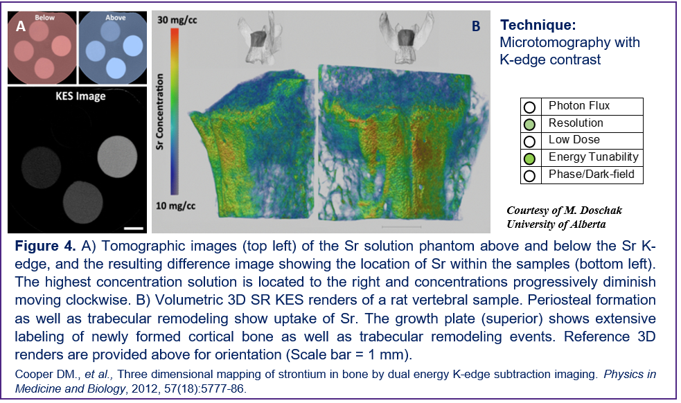
K-edge subtraction (KES) imaging was used as a non-invasive imaging technique to track the 3D distribution of strontium within animal models of bone disease, and to determine the impact of strontium incorporation in mineralized bone upon mineral density and bone strength.
Read more (bmit_highlights_jan2019.pdf)
Visualizing 3D Cytoarchitecture of the Human Cochlea in an Intact Temporal Bone
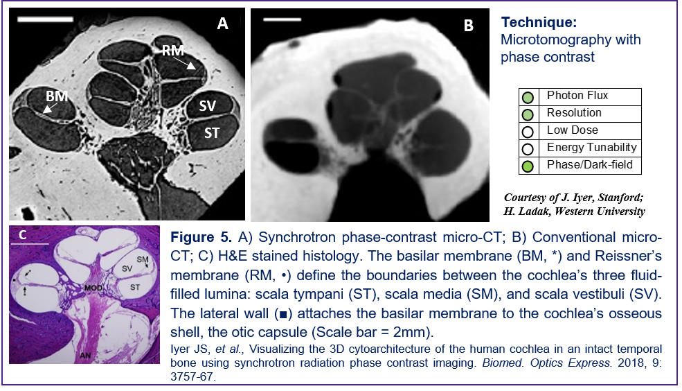
Posted on January 31, 2019
This panel shows the ability of synchrotron X-ray absorption and phase contrast imaging to enable visualization of sensory cells and nerve fibers in the cochlea’s sensory epithelium in situ (3D) in intact, non-decalcified, unstained human temporal bones.
Read more (bmit_highlights_jan2019.pdf)
Imaging Low-Contrast Scaffolds for Cardiac Regenerative Therapy
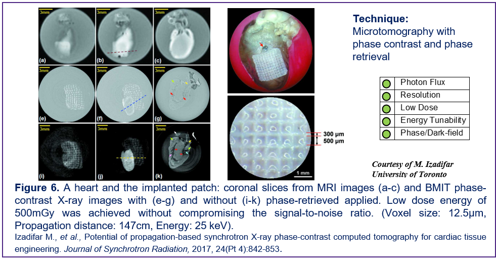
Posted on January 31, 2019
The visualization of hydrogel-based cardiac scaffolds is essential for longitudinal studies of cardiac regenerative therapy. Synchrotron X-ray phase contrast imaging computed tomography provides significantly more information than clinical MRI (3T) to visualize the microstructural features of the hydrogel cardiac patch for scaffold-based myocardial tissue engineering.
Read more (bmit_highlights_jan2019.pdf)
K-edge Subtraction Imaging of Barium and Strontium in Bone
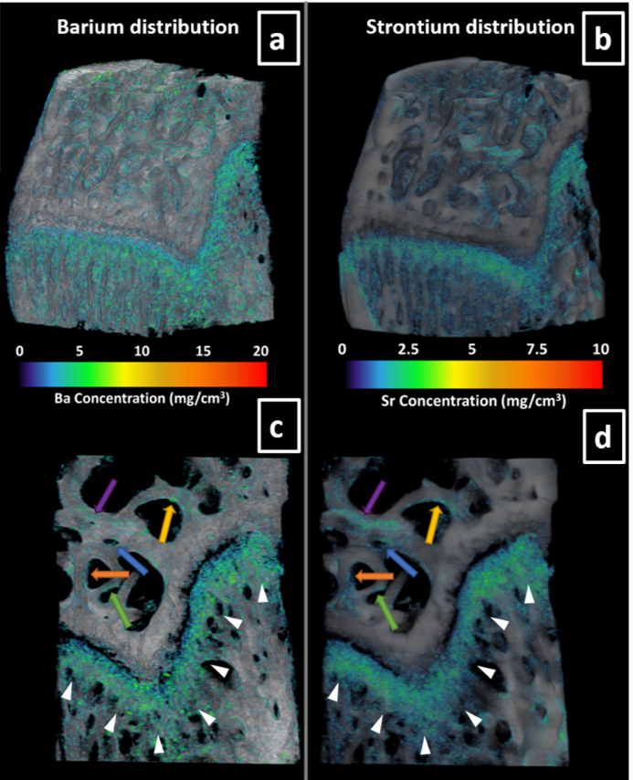
Posted on June 07, 2018
University of Saskatchewan researchers investigated skeletal uptake of barium and strontium, as tracers of bone turnover, in two different age groups, growing and skeletally mature, in healthy rats.
Read more (bmit-picture_of_the_month-june,_2018.pdf)
Studying model suspensions using high resolution synchrotron X-ray CT
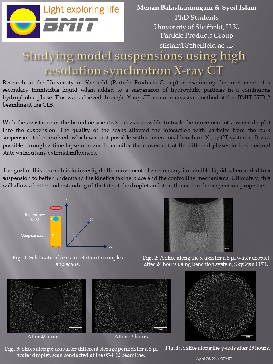
Posted on April 14, 2016
OIl and Water Researchers from the University of Sheffield used synchrotron X-ray CT to examine the fate of a water droplet in a suspension of sucrose and oil.
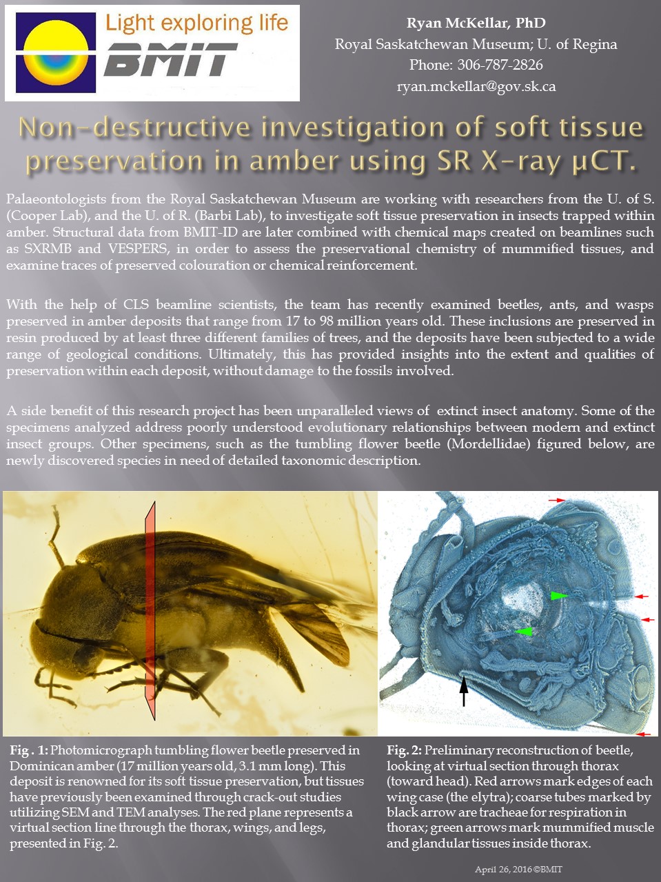
Non-destructive investigation of soft tissue preservation in amber
Posted on March 31, 2016
Preserved in Amber The plot of movies, soft tissue can be preserved in amber for many years. Paleontologists from the Royal Saskatchewan Museum are investigating soft tissue preservation of insects trapped within amber. The team has examined beetles, ants and wasps that range from 17 to 98 million years old in age.
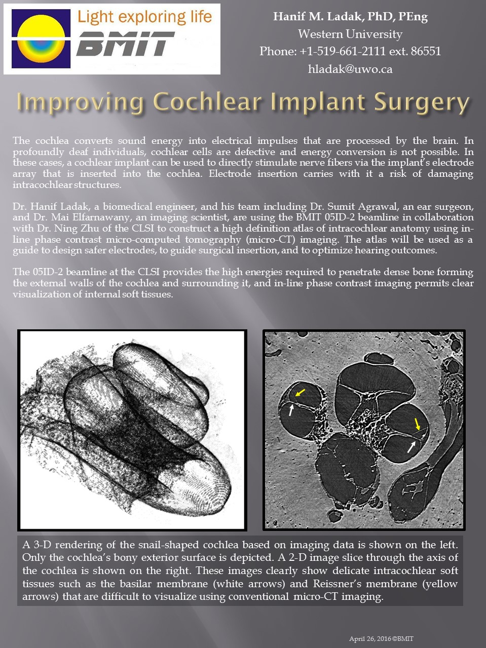
Improving Cochlear Implant Surgery
Posted on February 18, 2016
For a better sounding world A collaboration between Western University and the CLS to construct a high definition atlas of intracochlear anatomy using in-line phase contrast micro-computed tomography (micro-CT) imaging. The atlas will be used as a guide to design safer electrodes, to guide surgical insertion, and to optimize hearing outcomes.
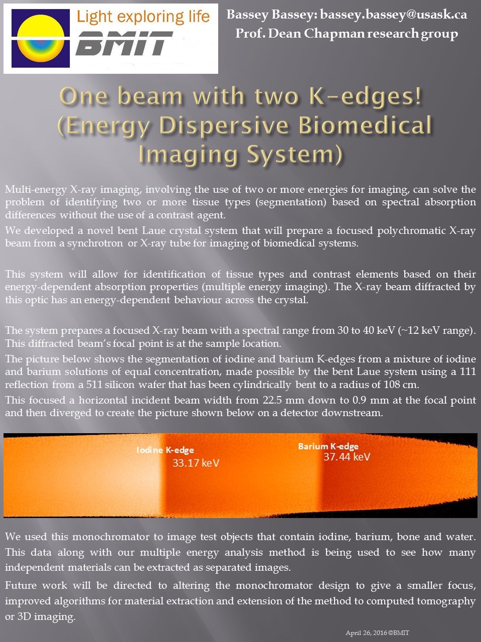
Energy Dispersive Biomedical Imaging System
Posted on November 30, 2015
One beam with two K-edges! Development has been done a multi-energy X-ray imaging system using a novel bent Laue crystal system. The focused beam hits the sample and then spreads out before reaching the detector. This results in an image where the horizontal relates to energy.
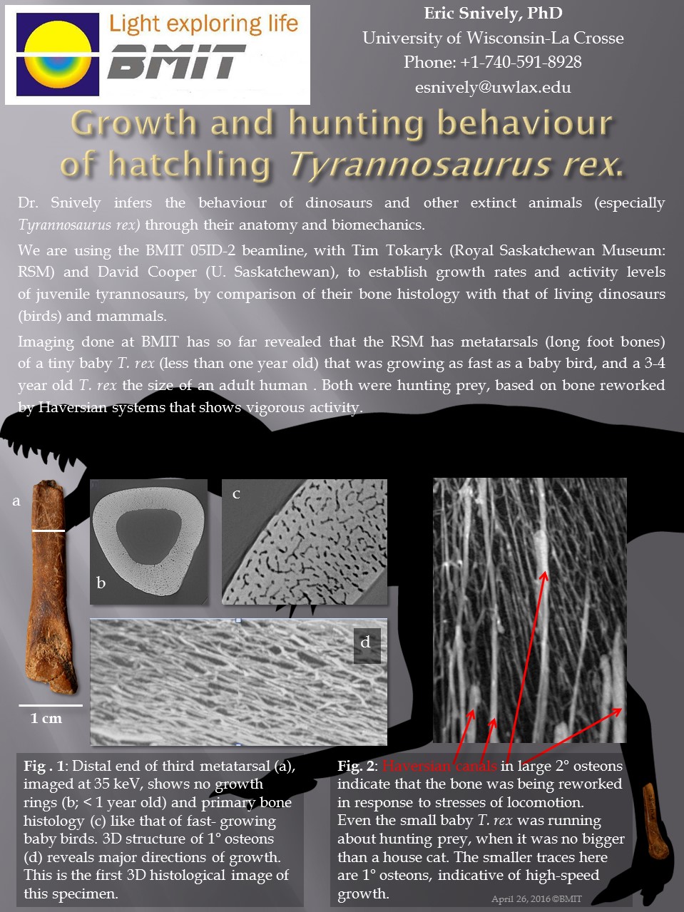
Growth and hunting behavior of hatchling Tyrannosaurus rex
Posted on September 08, 2015
Life of a baby dinosaur revealed Imaging of bones from young T.rex reveals insights into the lives of a hatchling (less than one year old) and a 3-4 year old T. rex the size of an adult human. The hatching was growing as fast as a baby bird and both were hunting prey showing the vigorous activity of youth.
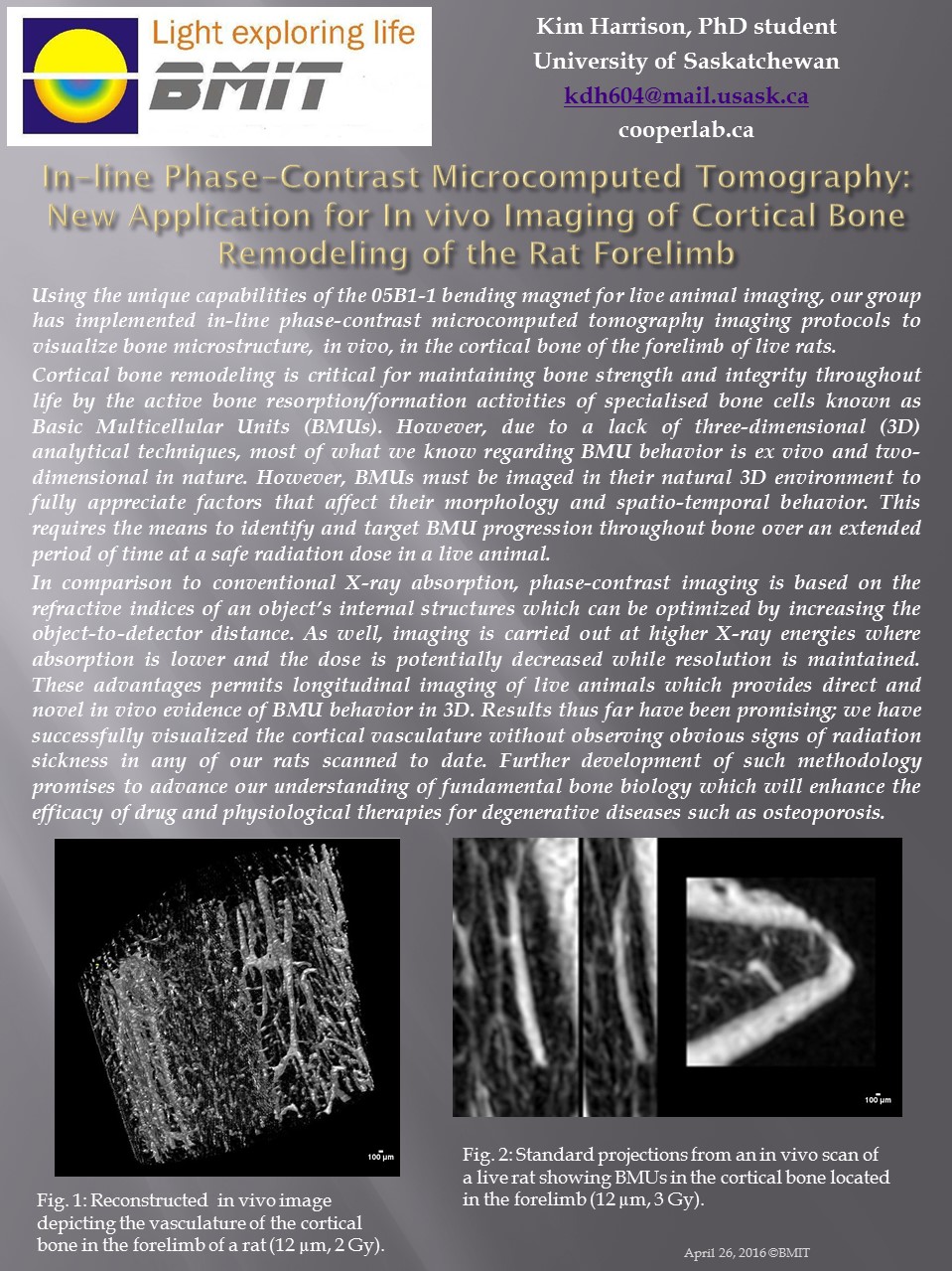
In-line Phase-Contrast Microcomputed Tomography
Posted on July 29, 2015
New Application for In vivo Imaging of Cortical Bone Remodeling of the Rat Forelimb In-line phase contrast was used to image cortical bone. Reducing the dose permits longitudinal imaging of live animals which provides direct and novel in vivo evidence of BMU behavior in 3D. Further development of such methodology promises to advance our understanding of fundamental bone biology which will enhance the efficacy of drug and physiological therapies for degenerative diseases such as osteoporosis.
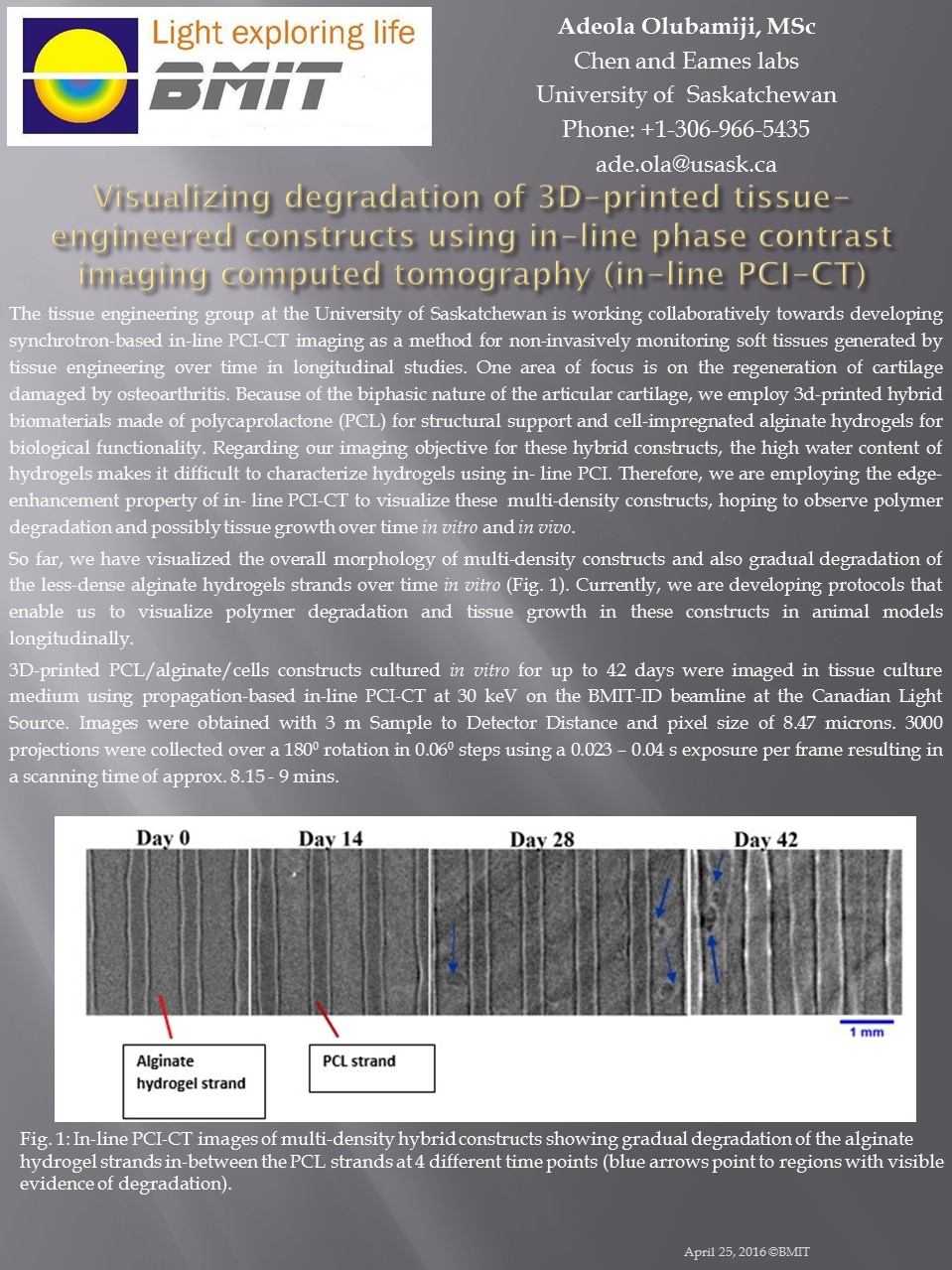
Visualizing degradation of 3D-printed tissue-engineered constructs
Posted on June 30, 2015
Using 3D printing to help grow new cartilage We employ 3d-printed hybrid biomaterials made of polycaprolactone (PCL) for structural support and cell-impregnated alginate hydrogels for biological functionality. Imaging is done using in-line phase contrast imaging computed tomography (in-line PCI-CT).
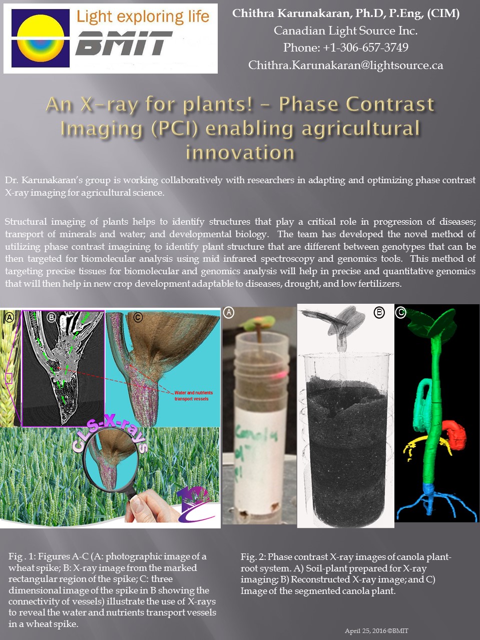
Phase Contrast Imaging (PCI) enabling agricultural innovation
Posted on May 29, 2015
An X-ray for plants! The team has developed a novel method of utilizing phase contrast imaging to identify plant structures. This is useful for studying the progression of disease, transport of minerals and water, and developmental biology.
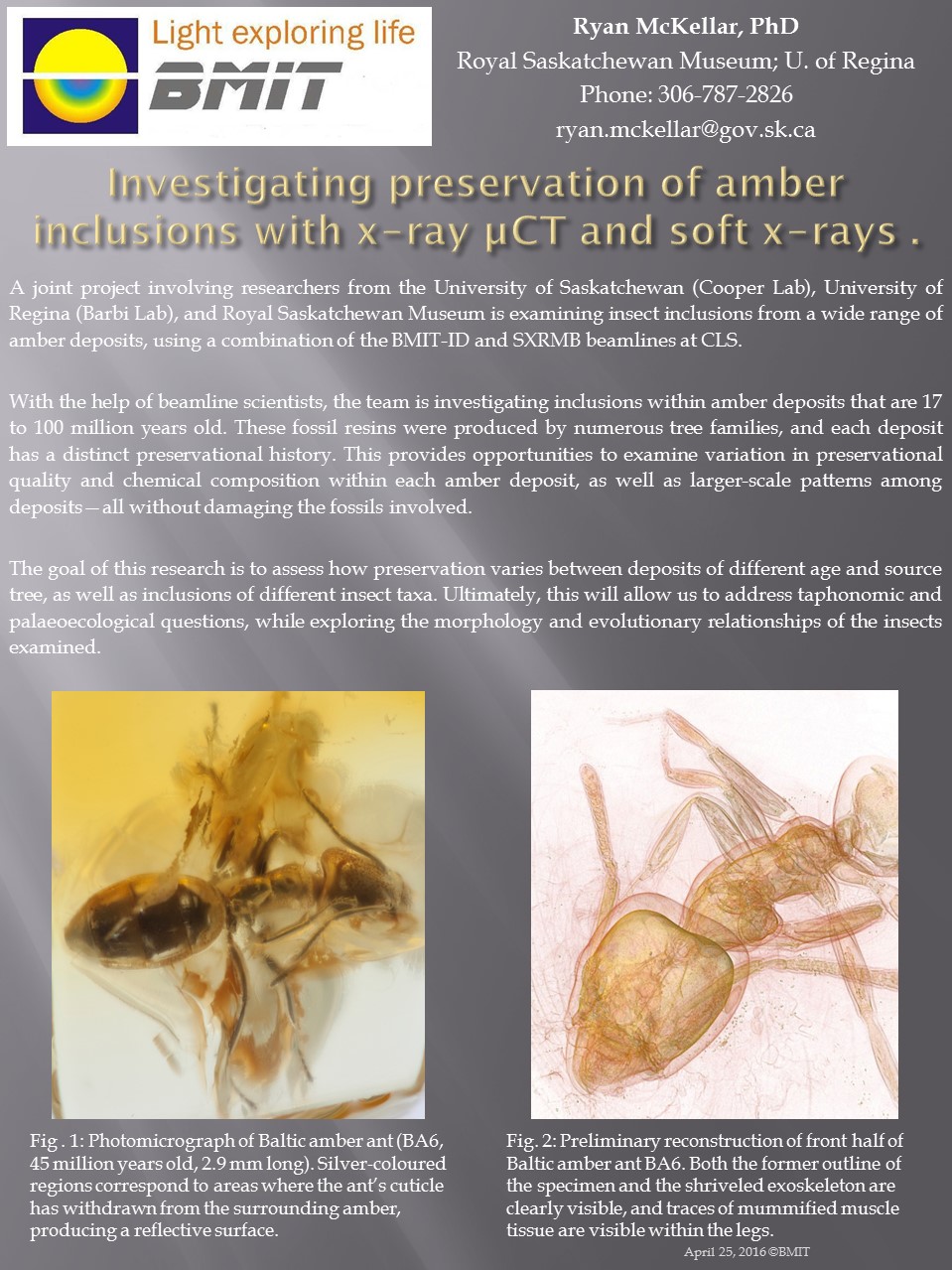
Investigating preservation of amber inclusions with X-ray µCT and soft X-rays
Posted on April 17, 2015
Frozen in time The team is investigating inclusions within amber deposits that are 17 to 100 million years old. The goal is to assess how preservation varies between deposits of different age and source tree.
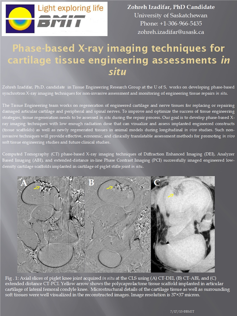
Phase-Based X-Ray Imaging Techniques for Cartilage Tissue Engineering Assessments in Situ
Posted on March 31, 2015
Repairing nerves and cartilage The tissue engineering team at the University of Saskatchewan is working on artificial tissue which will allow for the repair of cartilage and nerve tissue.
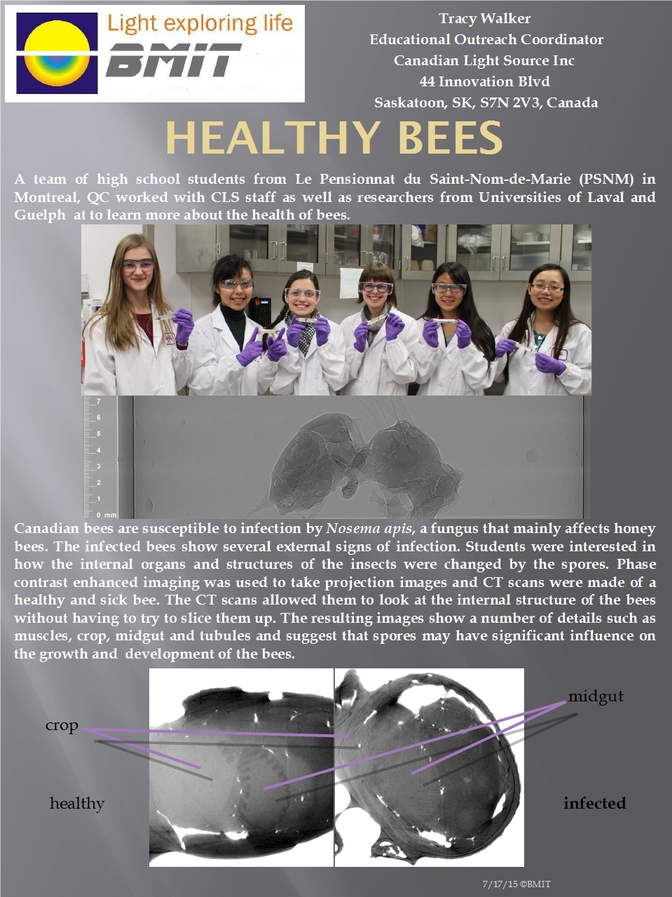
Healthy Bees
Posted on January 31, 2015
How fungus changes bees High school students from Quebec visited BMIT to image honey bees- comparing healthy bees to those infected by a fungus which targets Canadian honey bees.
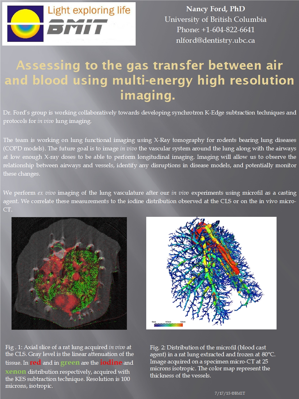
Assessing the Gas Transfer between Air and Blood Using Multi-Energy High Resolution Imaging
Posted on May 31, 2014
How diseased lungs are different The Ford group likes to watch rats breathe, comparing the relationship between airways and blood vessels in rats with pulmonary diseases.
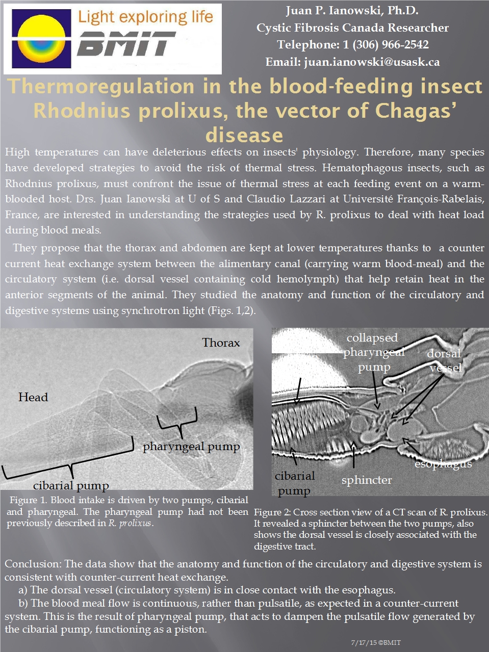
Thermoregulation in the Blood-Feeding Insect Rhodnius prolixus, the Vector of Chagas’ disease
Posted on April 30, 2014
Helping prevent the spread of disease Researchers from Saskatchewan and France are learning about how this insect pumps our hot blood through its cold body without dying. Understanding the mechanism with which the insect pumps our blood will help us combat its nefarious disease-spreading activities.
Beam Expander Update
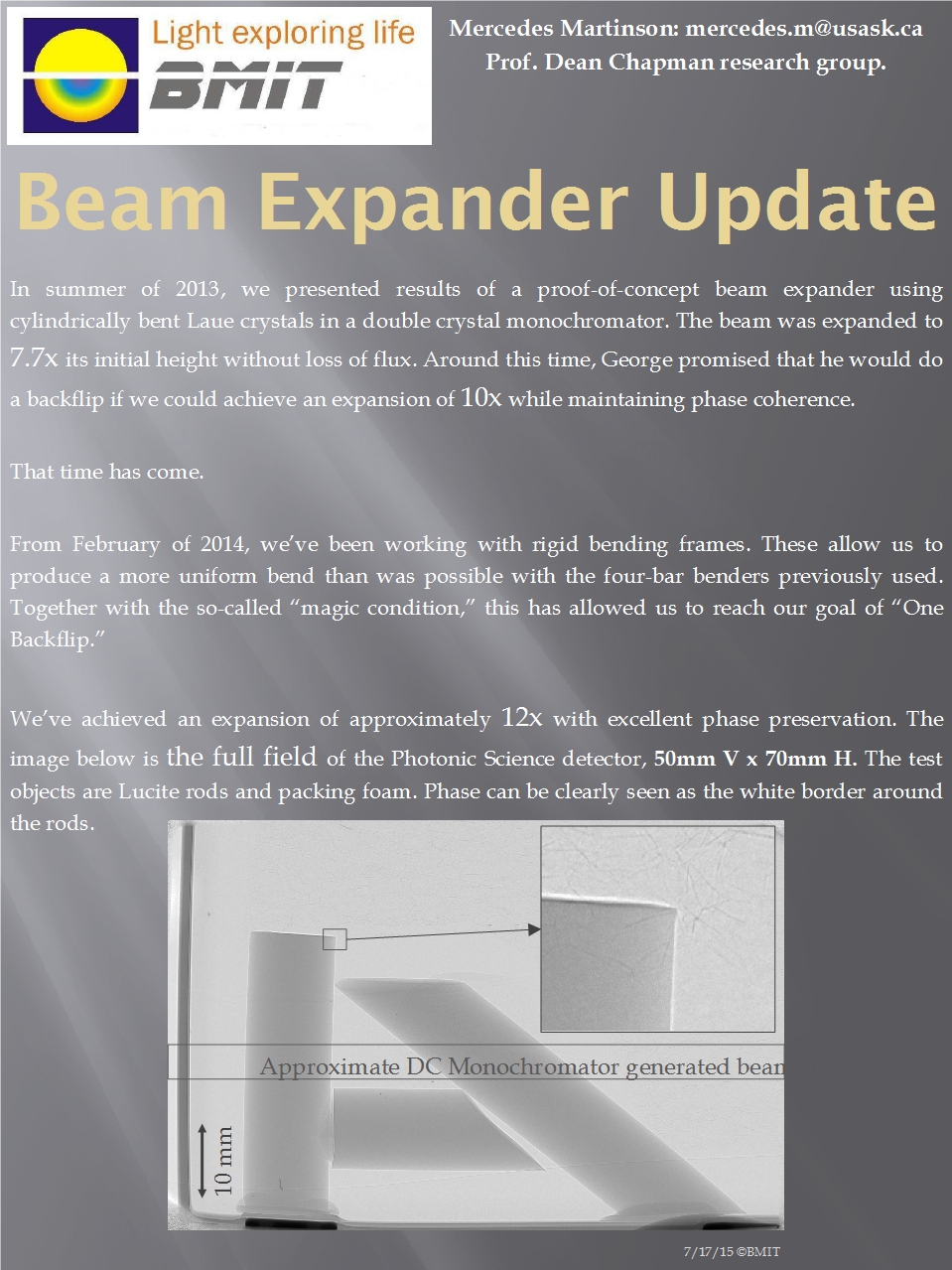
Posted on February 28, 2014
BMIT- bigger and better With much ingenuity, the available height of a BMIT image has been increased tenfold without ANY loss of flux or phase coherence. George almost died.
Fangtooth - Deep Sea Fish Skeleton
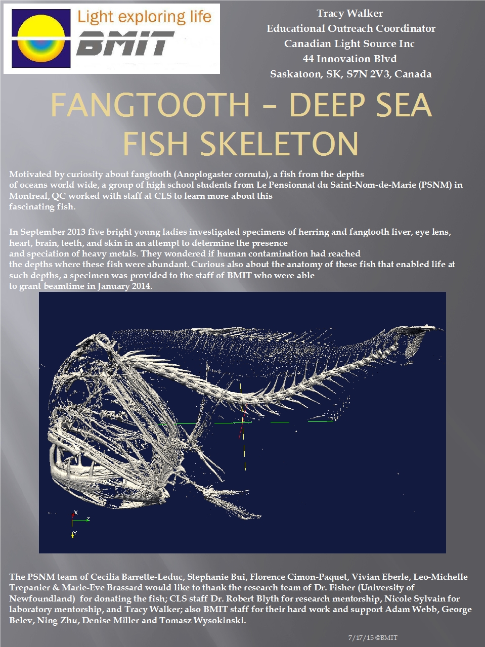
Posted on January 31, 2014
Fangtooth helps in determination of oceanic pollutants A group of high school students from Montreal scanned our fanged friend to figure out whether it harbors significant levels of various pollutants, which would suggest that we humans have managed to spread our waste pretty much everywhere on earth.
DEI Breath Hold Imaging Of Mouse Lungs
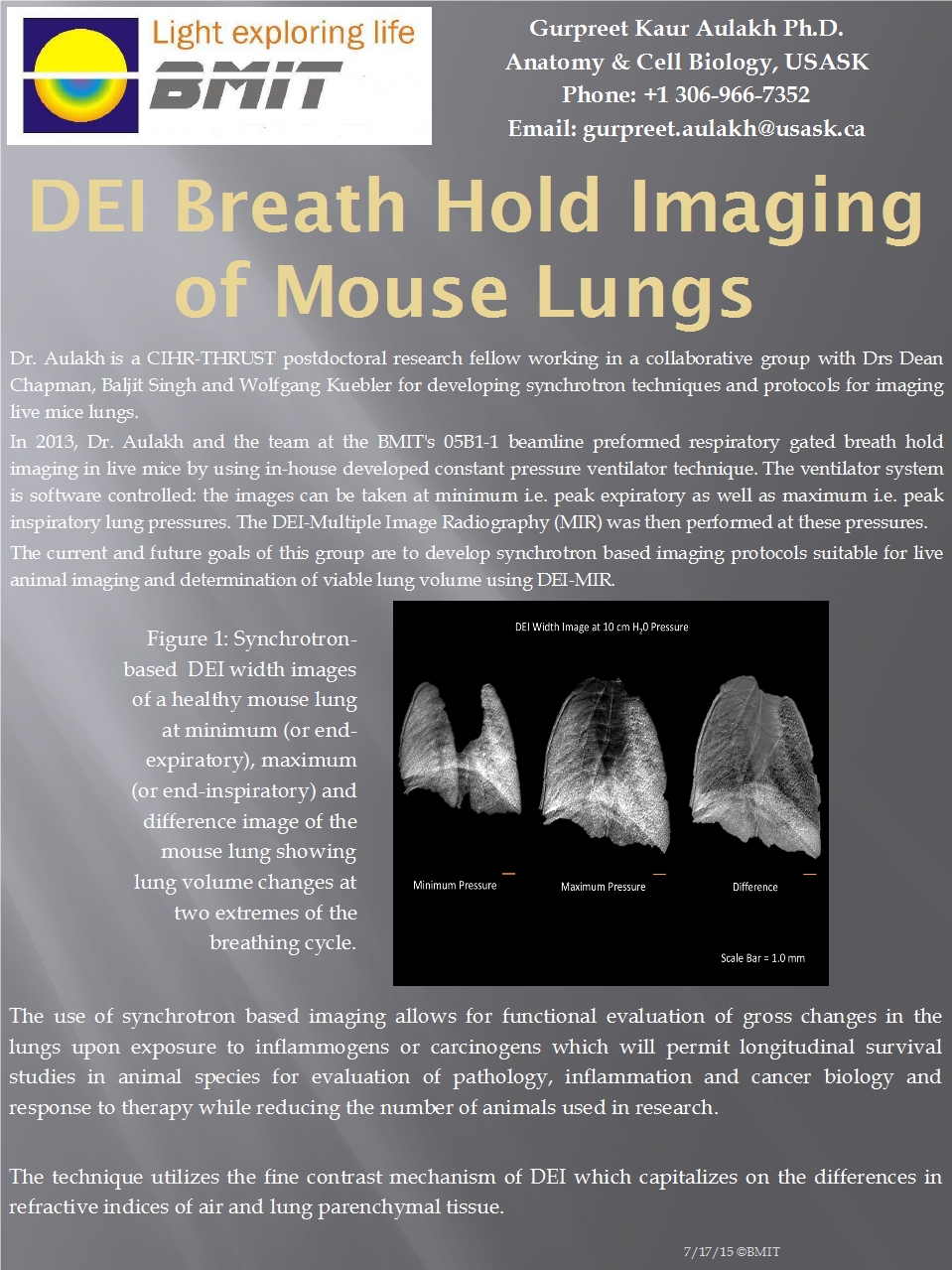
Posted on November 30, 2013
Helping animals breathe Software has been developed for imaging the lungs of live animals with synchrotron radiation. The software can be used to take pictures of the lungs at animals' maximum and minimum lung capacities, then to subtract the images from one another to see the difference. Seeing the difference between the maximum and minimum images while the animals are still alive allows researchers to better understand lung diseases and animals' responses to various treatments.
Grating Based Talbot Interferometer Imaging
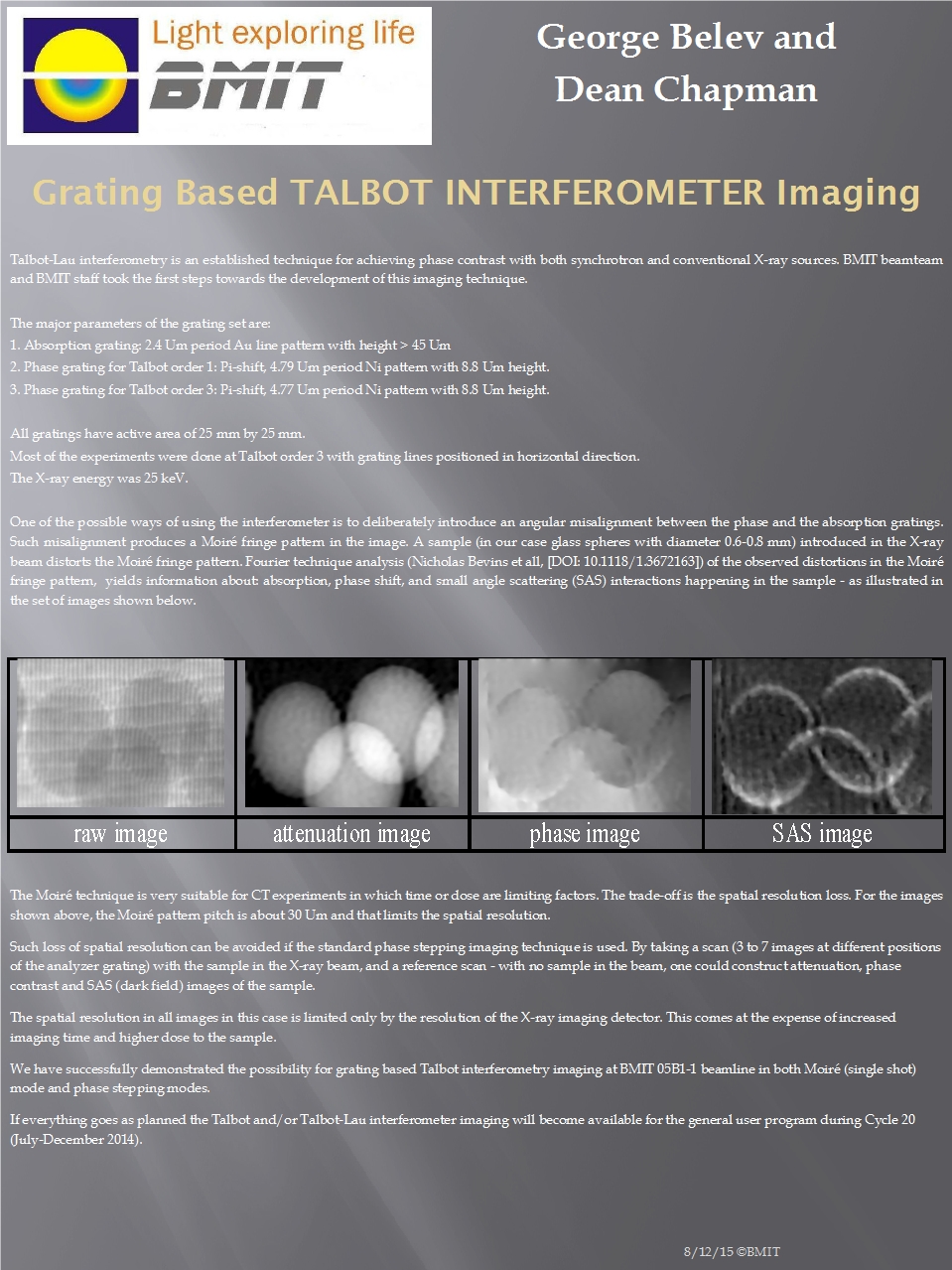
Posted on October 31, 2013
We’re working on it It should be ready for users by July 2014.
Beam Expander
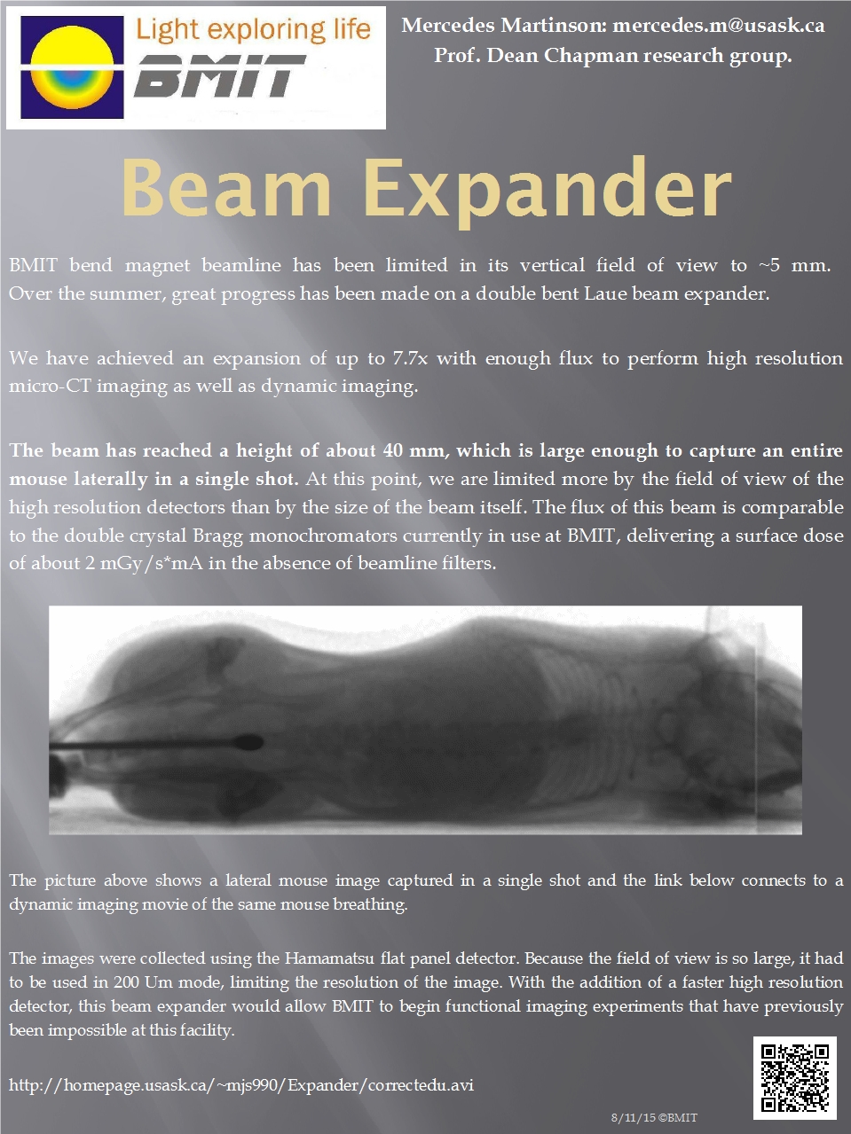
Posted on September 30, 2013
BMIT can image giants Using a double bent Laue beam expander, a mouse can now be imaged laterally in a single shot.
One Beam With Two K-Edges!
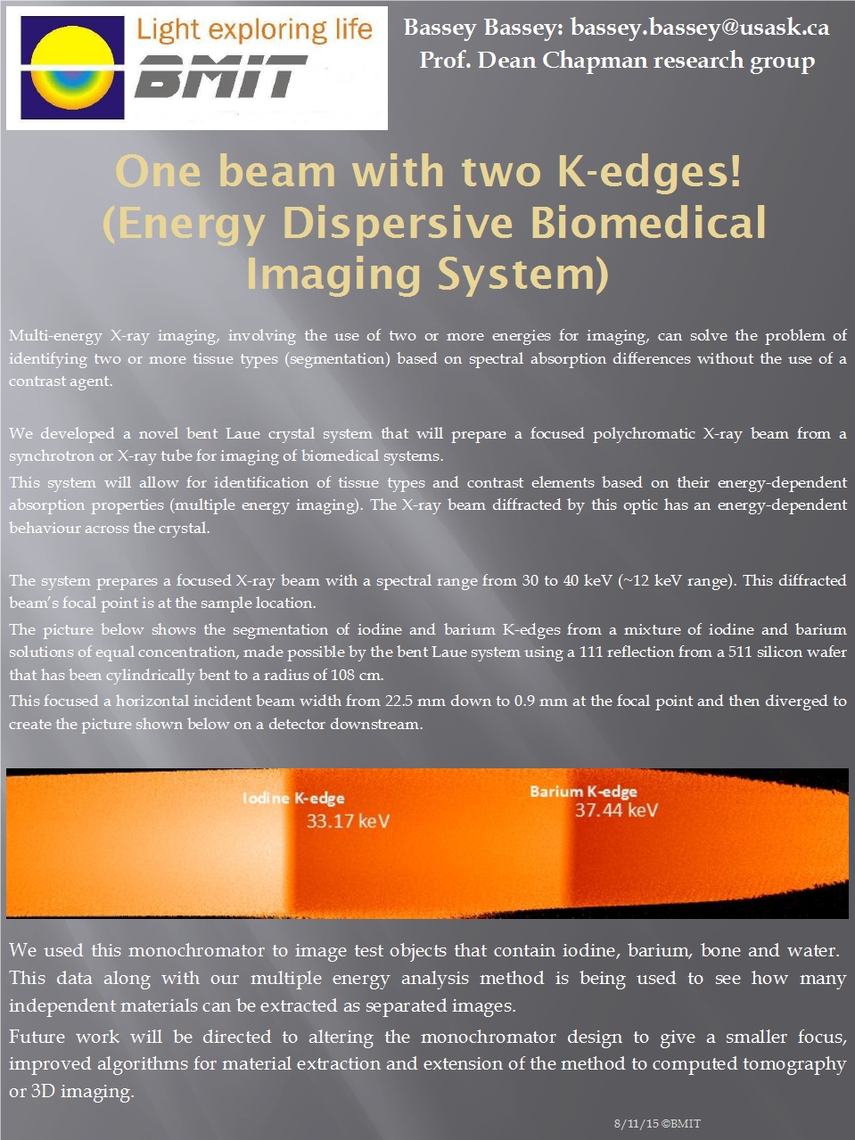
Posted on August 31, 2013
BMIT innovation Usually, if you have squares and circles in a sample, you have no choice but to look at an image which includes both the squares and the circles (superimposed upon one another- making it difficult to see anything useful). With two K-edges, you can have two completely different images- one with only squares, and one with only circles.
Soft Tissue Imaging Of the Temporomandibular Joint
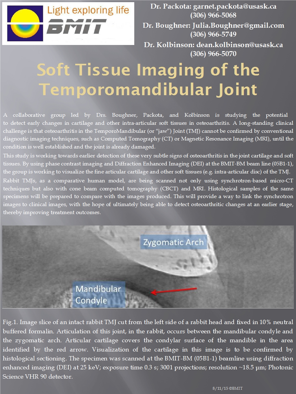
Posted on July 31, 2013
Helping diagnose arthritis of the jaw Using standard hospital equipment, arthritis of the jaw can’t be detected until it’s too late... BMIT can help.
Synchrotron Imaging Of the Spatial Distribution of Barium in Bone to Understand the Role of Remodeling In Bone Disease
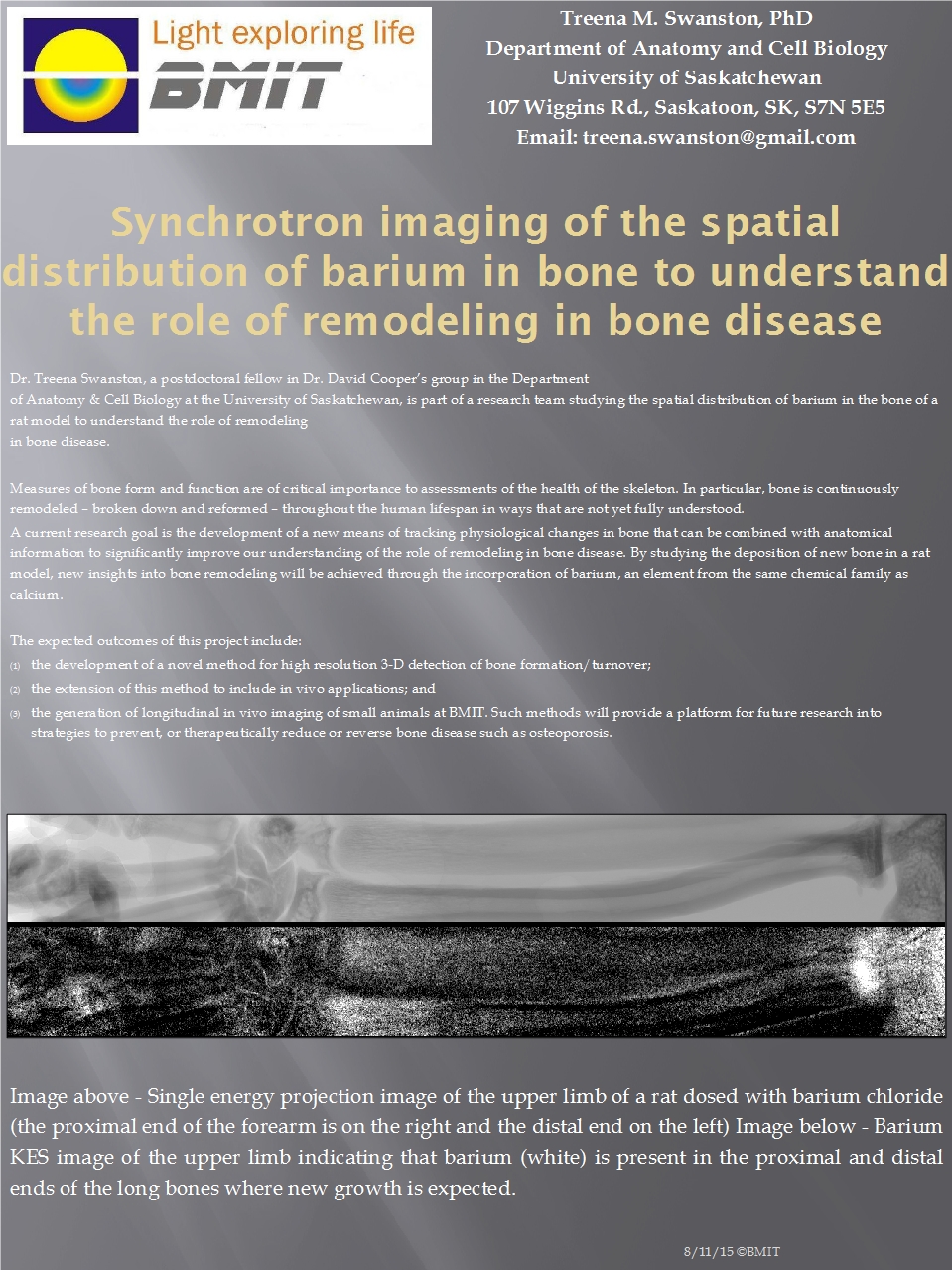
Posted on June 30, 2013
Your body treats your bones the way a kid treats Lego Your bones are constantly being destroyed and replaced by your body. One research team is using BMIT to figure out exactly how that process of destructive creation is affected by diseases like osteoporosis.
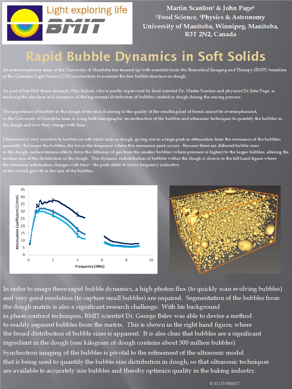
Rapid Bubble Dynamics in Soft Solids
Posted on May 31, 2013
The quest for the perfect loaf of bread Imaging techniques at BMIT are helping refine the standard industrial bread-baking quality assurance ultrasound technologies which are currently in use.
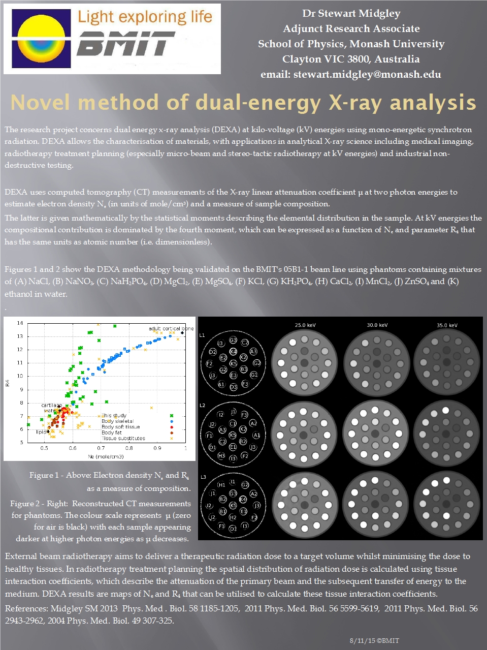
Novel Method of Dual-Energy X-Ray Analysis
Posted on April 30, 2013
Innovation at BMIT Dr. Midgley has been investigating DEXA for use in the characterization of materials, with applications in analytical x-ray science- including medical imaging, radiotherapy treatment planning, and industrial nondestructive testing.
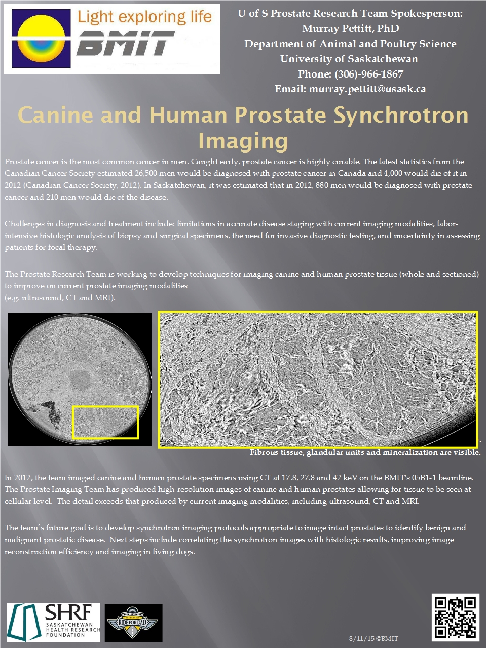
Canine and Human Prostate Synchrotron Imaging
Posted on March 31, 2013
Prostate cancer kills- but it doesn’t have to Caught early, prostate cancer is highly curable. The problem is catching it early. Using BMIT, a University of Saskatchewan research team is studying prostate cancer in dogs in an effort to apply what they learn to increasing the ability of currently available hospital technology to diagnose prostate cancer at an earlier stage.
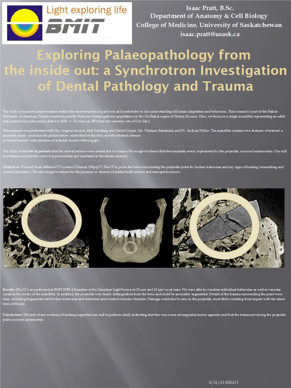
Exploring Paleopathology from the Inside Out: A Synchrotron Investigation of Dental Pathology and Trauma
Posted on February 28, 2013
Point taken: An unusual case of incisor agenesis and mandibular trauma in Early Bronze Age Siberia Synchrotron imaging was used to diagnose two unusual but unrelated conditions. The first is one of the earliest cases involving dental agenesis, the failure of two front teeth to develop. The second, the embedded projectile tip, represents an almost certain deliberate assault that occurred at or around the time of death.
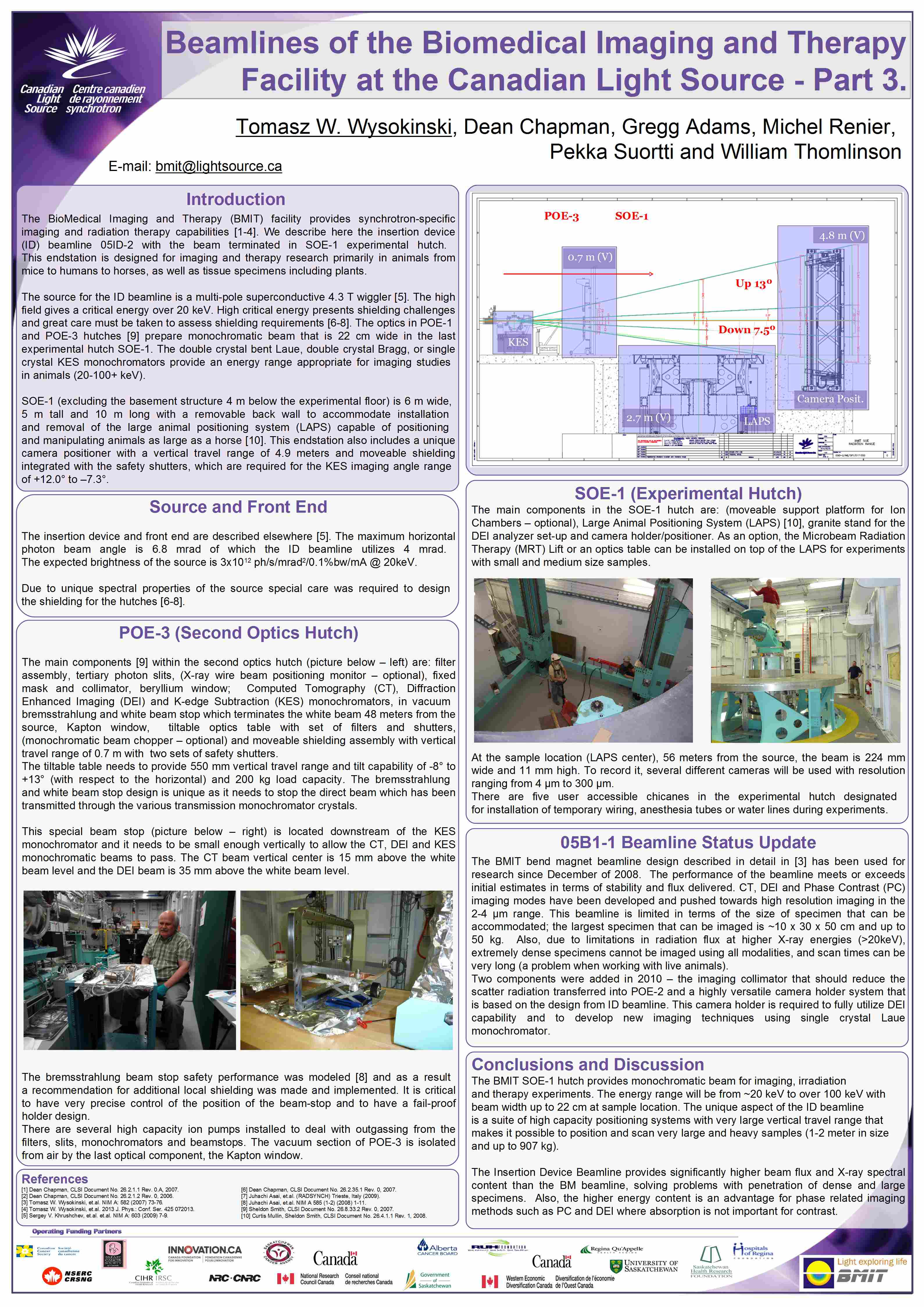
Beamlines of the Biomedical Imaging and Therapy Facility at the Canadian Light Source
Posted on January 01, 2013
05ID-2 overview We describe here the insertion device (ID) beamline 05ID-2 with the beam terminated in SOE-1 experimental hutch. This endstation is designed for imaging and therapy research primarily in animals from mice to humans to horses, as well as tissue specimens including plants.
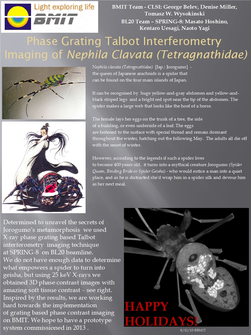
Phase Grating Talbot Interferometry Imaging of Nephila clavata (Tetragnathidae)
Posted on December 31, 2012
Spider Queen While imaging at SPRING-8, staff from BMIT were inspired to develop grating based Talbot interferometry.
Precision Bending of Silicon Wafers for Bent Crystal Monochromators
Posted on November 30, 2012
High quality mono beam for less Improving bent crystal monochromators and studying the utility of silicon wafers as bent crystal optic components.
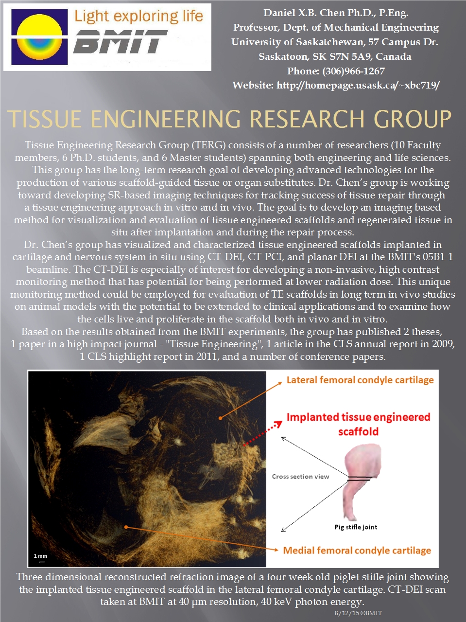
Tissue Engineering Research Group
Posted on October 31, 2012
Replacement body parts for all BMIT allows non-invasive monitoring of how well implanted tissue performs, and exactly where the implants are and/or are not healing.
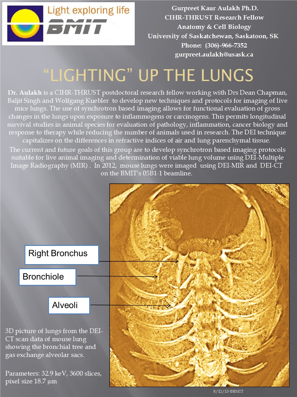
“Lighting” Up the Lungs
Posted on September 30, 2012
Development of lung imaging The goal of this work is to develop synchrotron based imaging protocols suitable for live animal imaging and for functional evaluation and measurement of gross changes in the lungs.
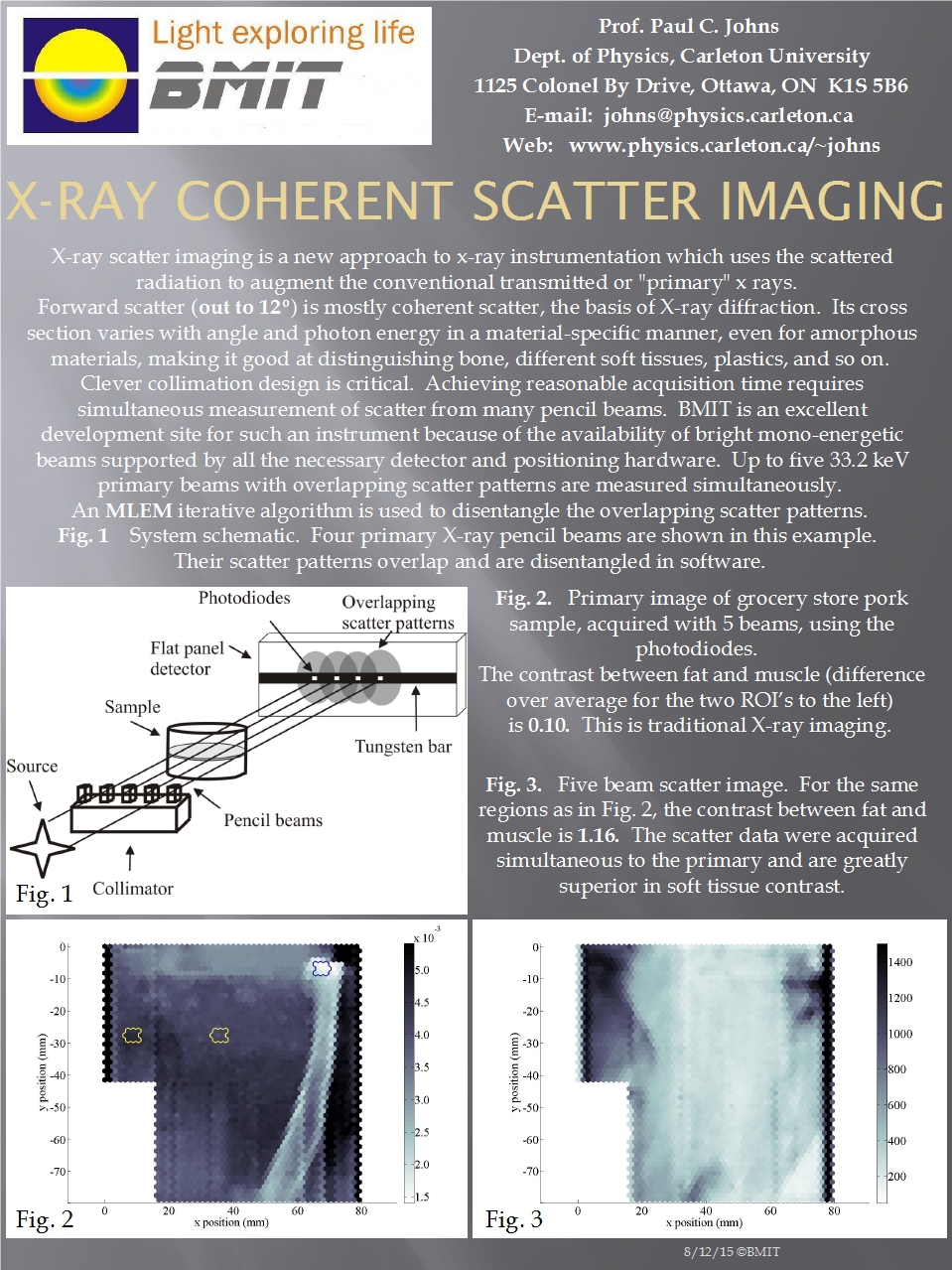
X-Ray Coherent Scatter Imaging
Posted on August 31, 2012
Extracting information from "random" scatter X-ray scatter imaging uses the scattered radiation to augment conventional X-ray imaging.
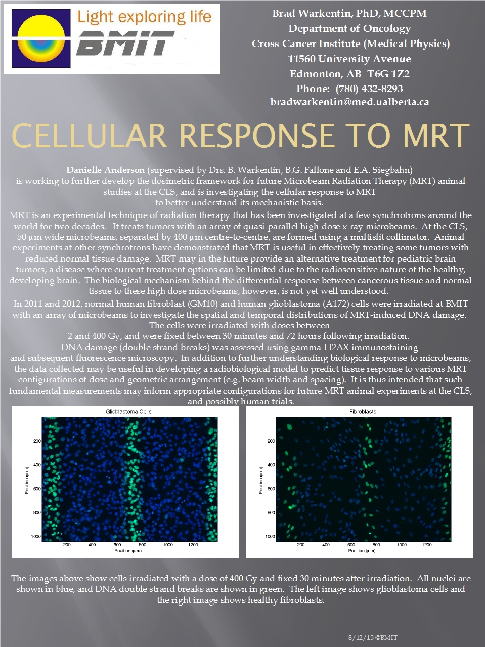
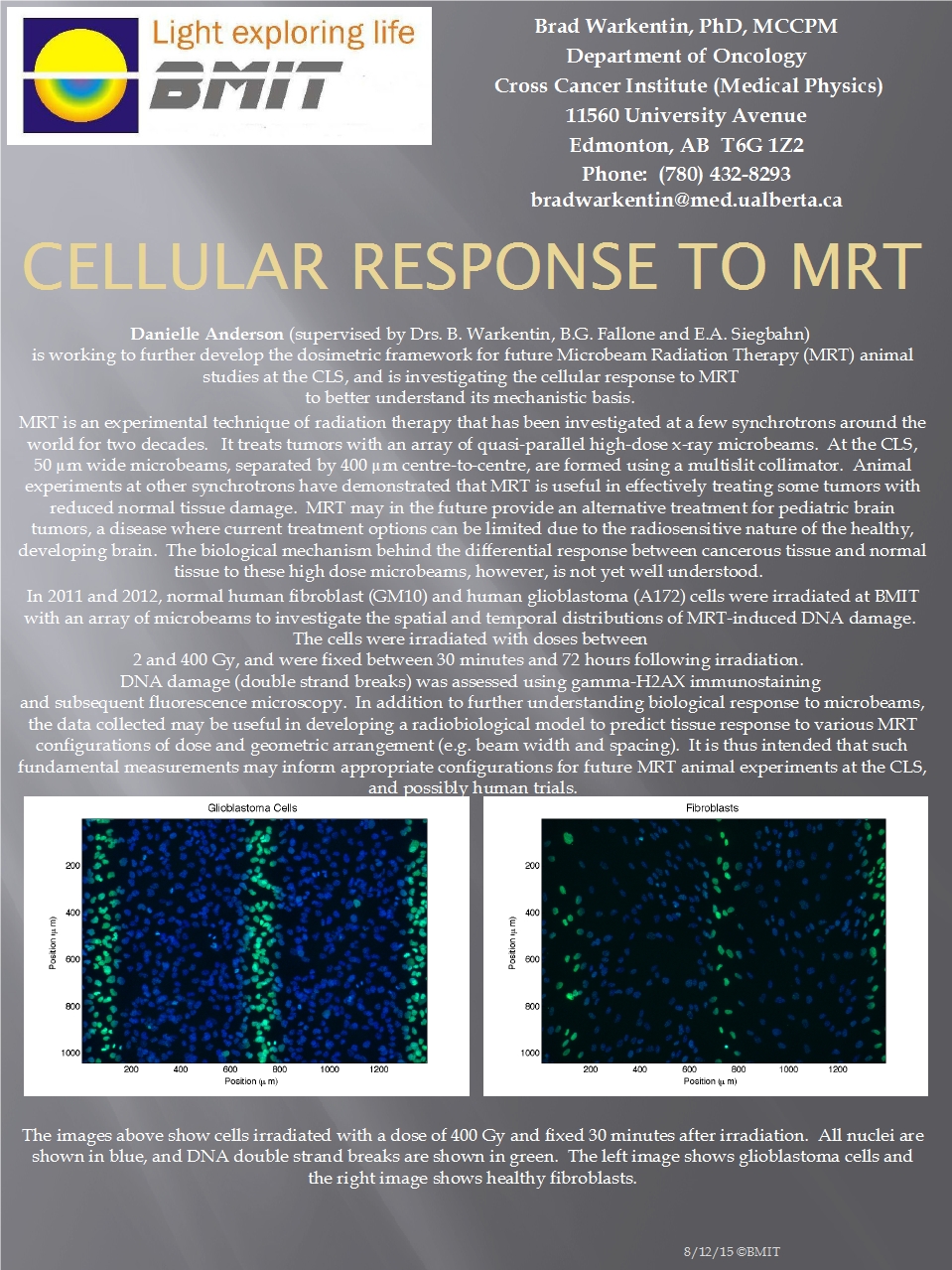
Cellular Response to Microbeam Radiation Therapy (MRT)
Posted on July 31, 2012
A precise radiation beam for cancer elimination A scientist from the University of Alberta is developing a method for using BMIT’s beam to kill cancerous tissue without harming the surrounding healthy tissue.
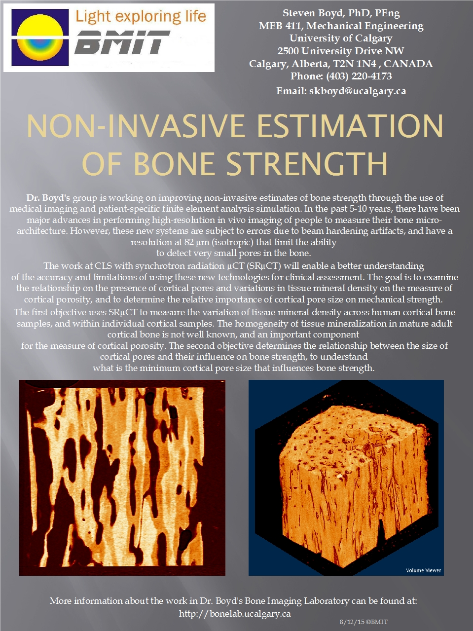
Non-Invasive Estimation of Bone Strength
Posted on June 30, 2012
Do the pores in your bones bode ill for your future? Dr. Boyd and his team may be able to tell you.
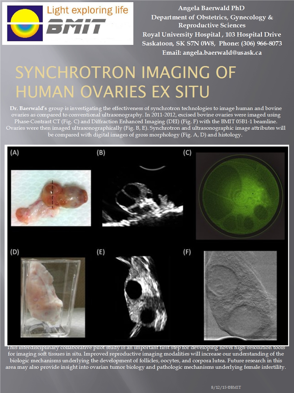
Synchrotron Imaging Of Human Ovaries ex situ
Posted on May 31, 2012
The business of helping life come into this world is both messy and beautiful Improving soft tissue imaging, understanding ovarian cancer, and figuring the mechanisms through which infertility may occur.
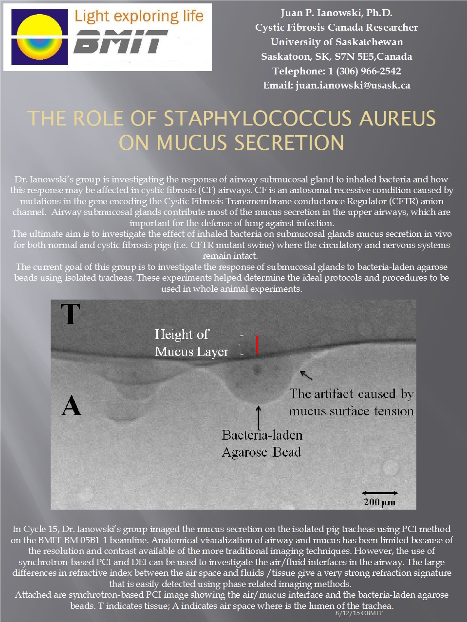
The Role of Staphylococcus Aureus on Mucus Secretion
Posted on April 30, 2012
How cystic fibrosis affects the mucus protecting us from pulmonary infection. Response of the airway submucosal gland to inhaled bacteria with and without cystic fibrosis.
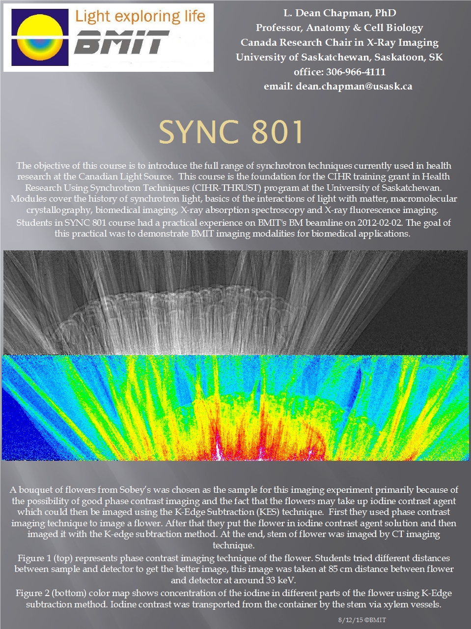
SYNC 801
Posted on March 31, 2012
A class about the application of synchrotron radiation to problems in the life sciences Covering the history of synchrotron light, interactions of light with matter, macromolecular crystallography, biomedical imaging, x-ray absorption imaging, and x-ray flurorescence imaging.
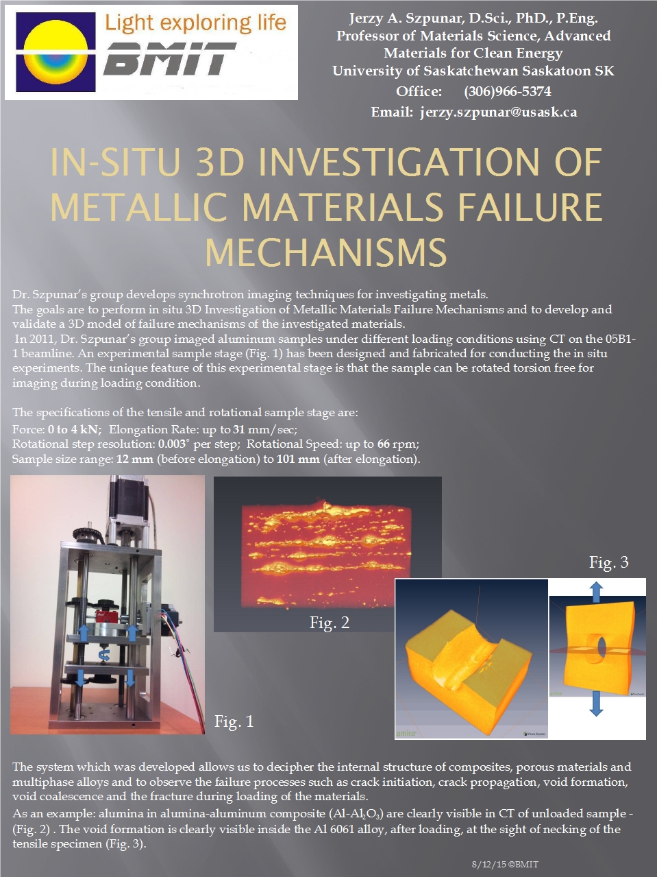
In-situ 3D Investigation of Metallic Materials’ Failure Mechanisms
Posted on February 29, 2012
Measuring failure The bright x-rays of BMIT have allowed the Szpunar group to observe the failure of various materials at the micron level.
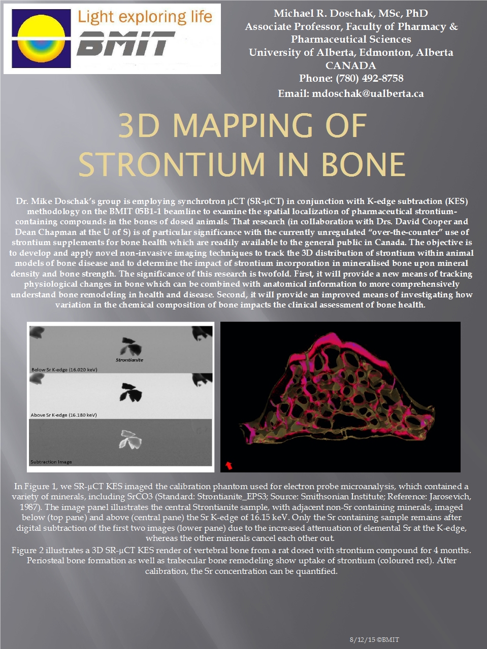
3D Mapping Of Strontium in Bone
Posted on January 31, 2012
Strontium bone-strength supplements? We don't know exactly how useful strontium supplements are, but researchers are in the process of figuring it out. They feed rats strontium, and then watch how concentrations of the element in the rats' bones change over the rats' lives using micro Computed Tomography (uCT) and K-Edge Subtraction (KES) techniques.
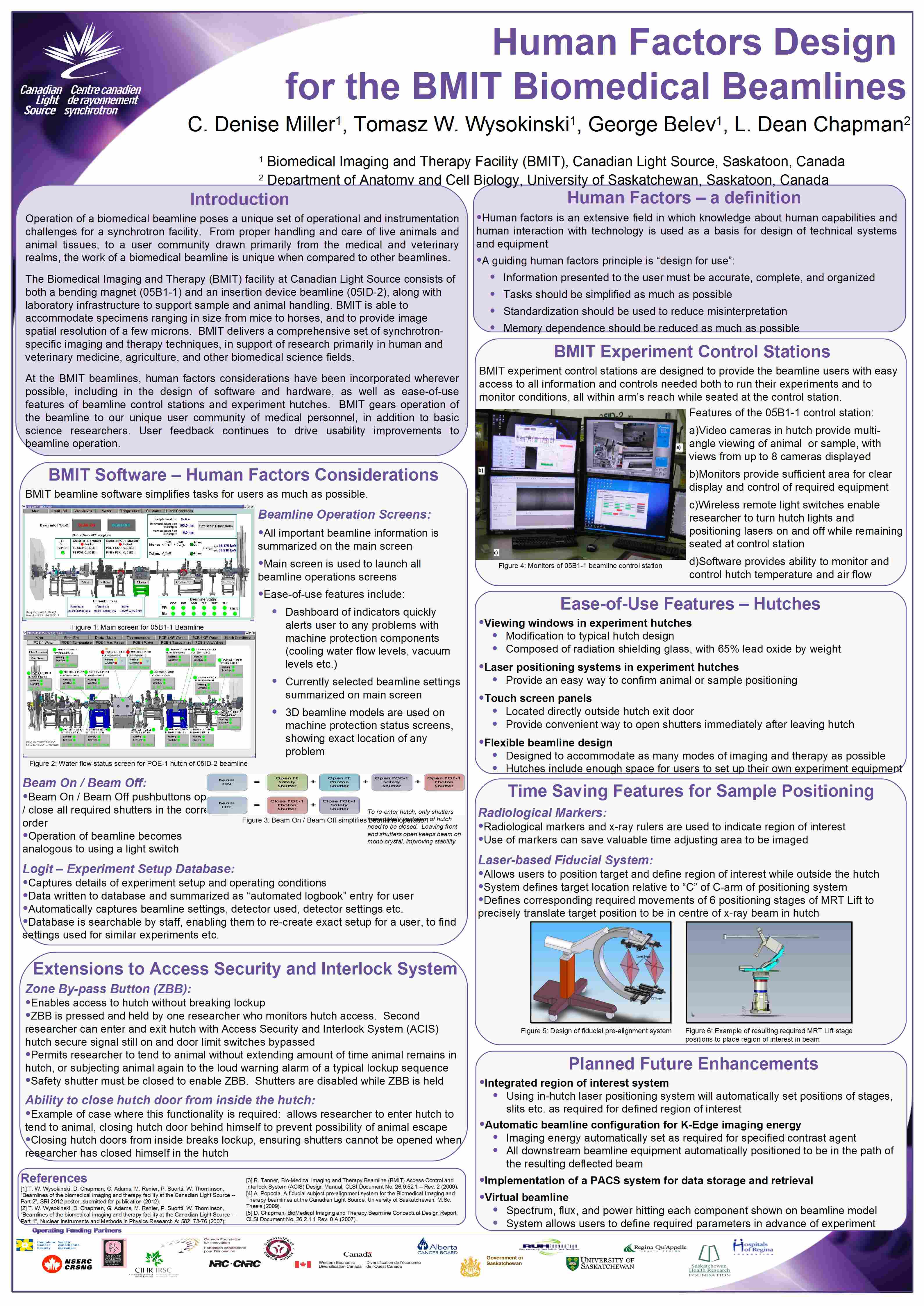
Human Factors Design for the BMIT Biomedical Beamlines
Posted on January 01, 2012
BMIT designed for ease of use At the BMIT beamlines, human factors considerations have been incorporated wherever possible, including in the design of software and hardware, as well as ease-of-use features of beamline control stations and experiment hutches. BMIT gears operation of the beamline to our unique user community of medical personnel, in addition to basic science researchers. User feedback continues to drive usability improvements to beamline operation.
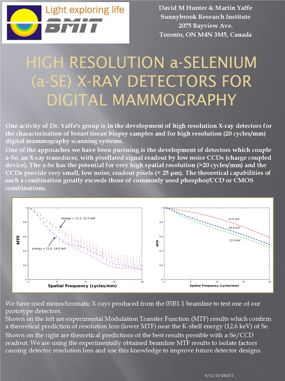
High Resolution a-Selenium (a-Se) X-Ray Detectors for Digital Mammography
Posted on November 30, 2011
Towards higher resolution mammography A prototype detector was tested in the development of high resolution detectors for digital mammography.
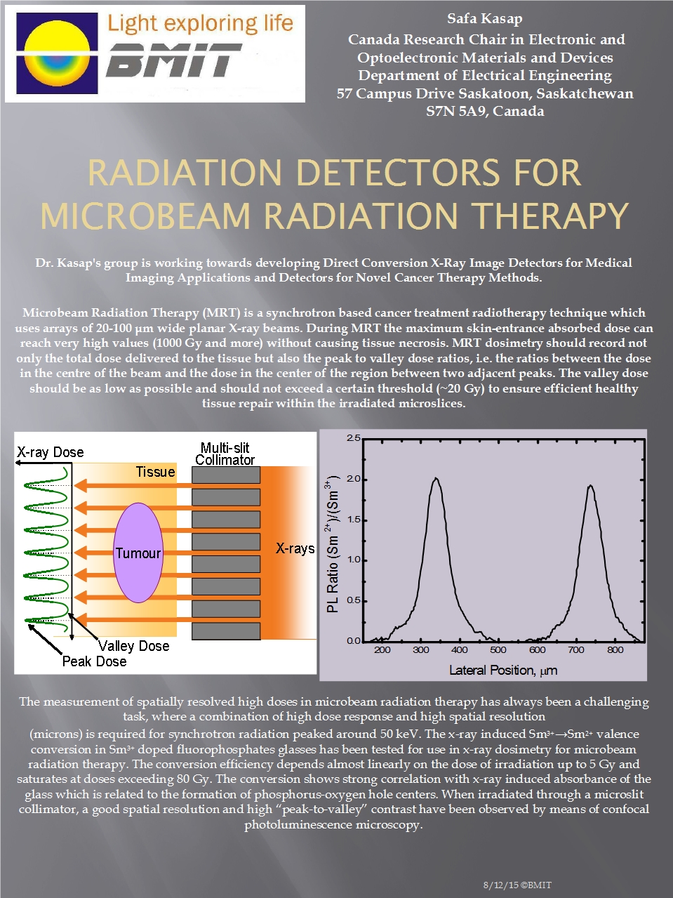
Radiation Detectors for Micro-Beam Radiation Therapy
Posted on October 31, 2011
Precision dose measurements for radiation therapy The measurement of spatially resolved high doses in micro-beam radiation therapy is a challenging task requiring the combination of high dose response and high spatial resolution.
Insect Flight Muscle Phase Polymorphism
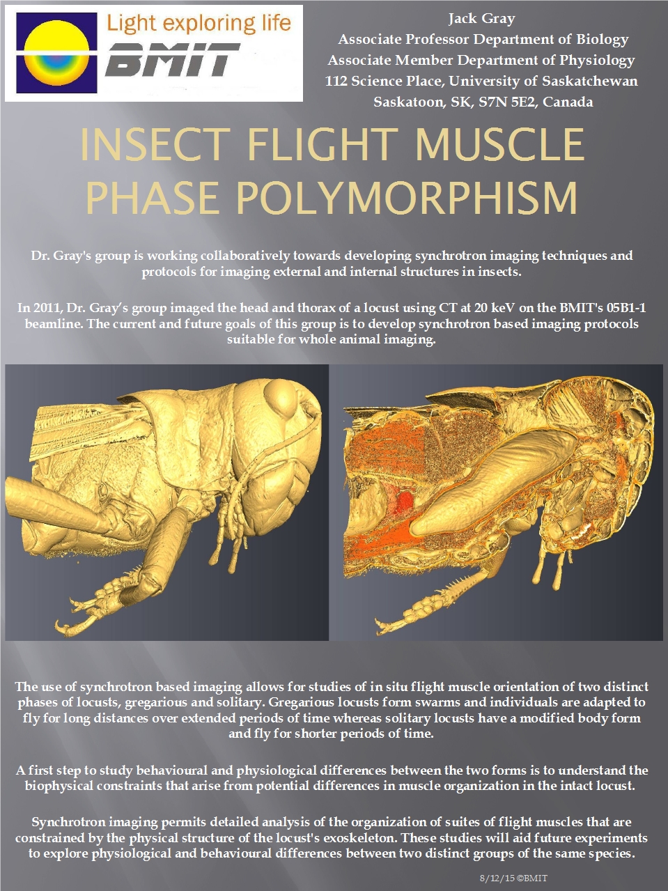
Posted on September 30, 2011
Why are some locusts so lonesome? A research group at the University of Saskatchewan is using BMIT to develop an understanding of how the exoskeletons of two different groups within the same species of locust constrain the development of their flight muscles.
DEI Imaging of Growing and Inflamed Joints
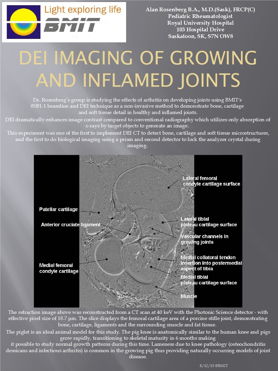
Posted on August 31, 2011
Fighting early-onset arthritis The Rosenberg group is breaking new ground in the study of joint disease.
Investigating Clinical Applications of Synchrotron Imaging In Veterinary Medicine
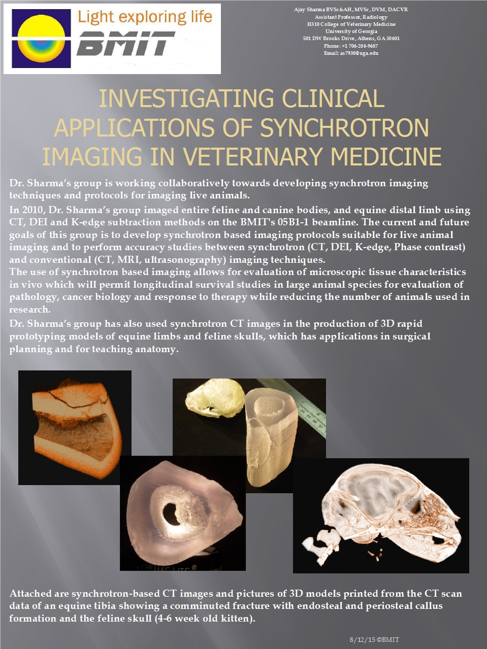
Posted on July 31, 2011
Longitudinal survival studies with large animals The Sharma group is using BMIT for comparative analysis, surgical planning, live animal imaging, and building 3D teaching models.
Microfluidic and Nanofluidic Transport Phenomena
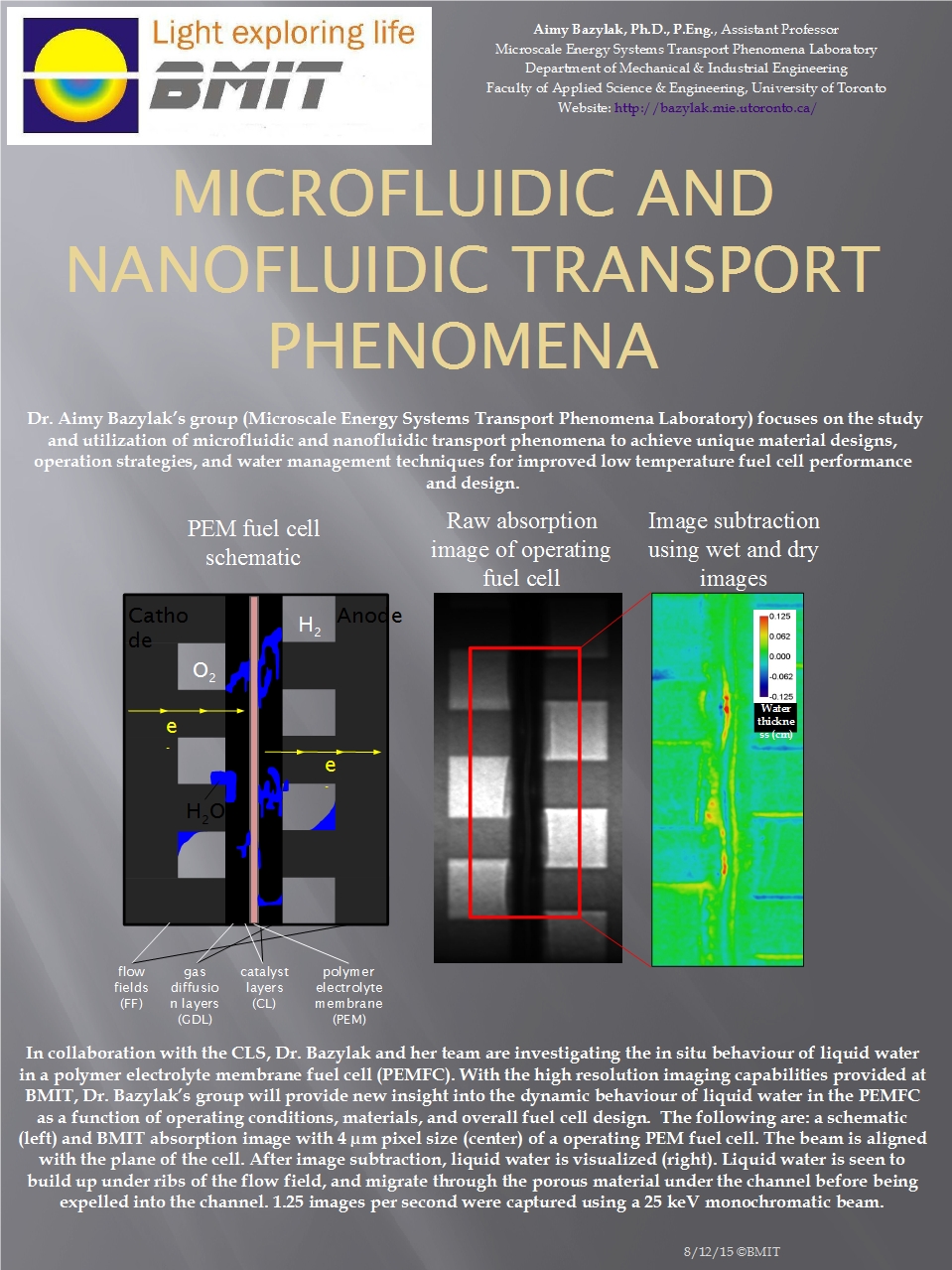
Posted on June 30, 2011
Understanding fluid flow at the micro- and nano-scale Measuring the behavior of liquid water in a fuel cell provides new insights into the function of fuel cells and may lead to improvements in materials and design for low temperature fuel cells.
Measuring the Biomechanics of Normal and Pathological Joints
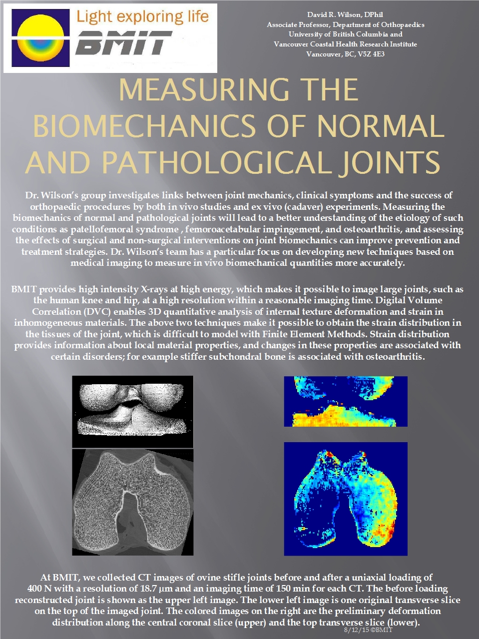
Posted on May 31, 2011
High resolution imaging of joints Dr. Wilson's group is helping improve diagnoses of joint problems by conducting stress analyses in the BMIT beam.
Imaging Embryonic Tooth Development in Mouse
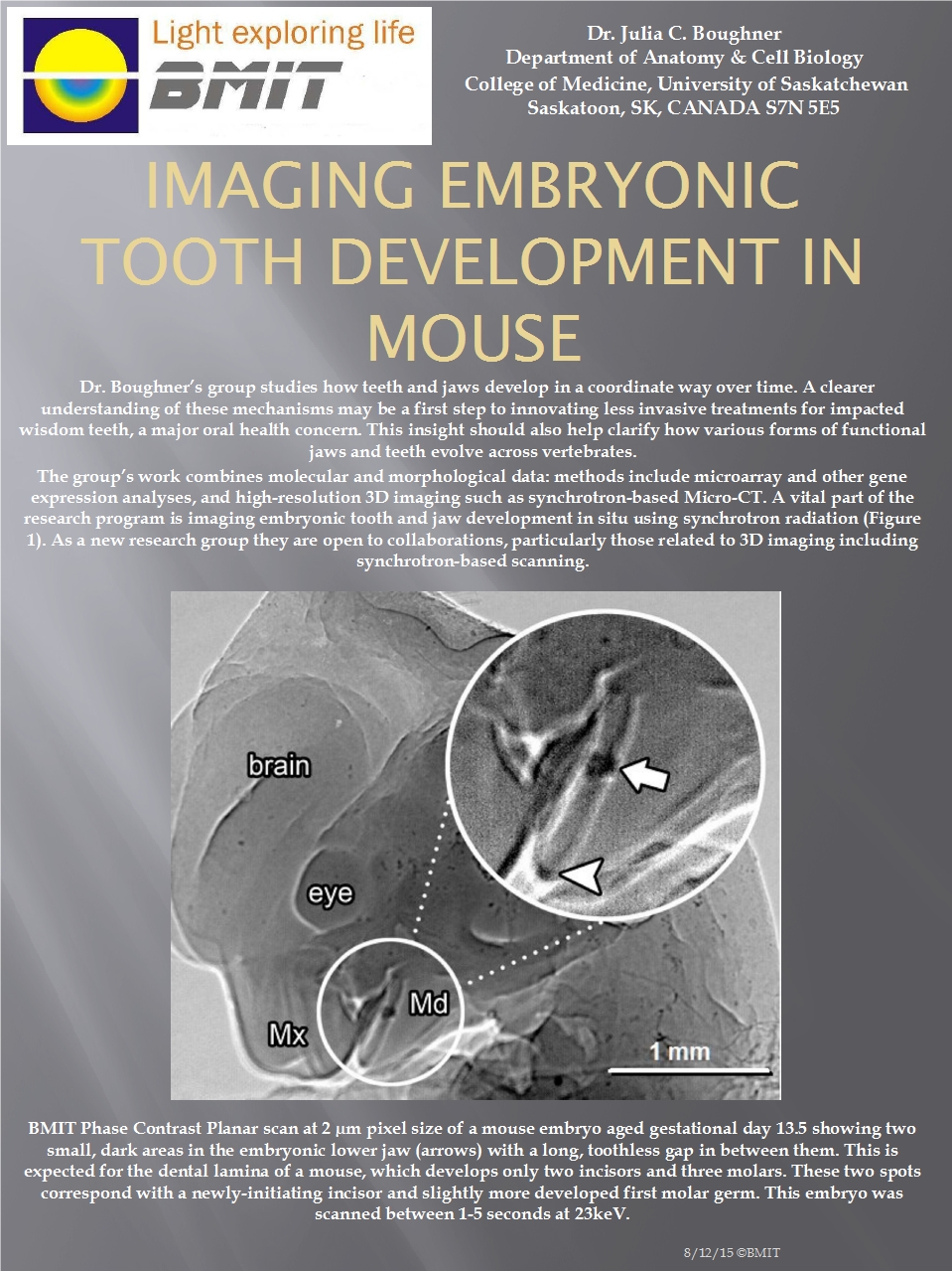
Posted on April 30, 2011
The teeth of mice and men Studying how teeth and jaws develop in a coordinated way over time.
High Resolution 3D Imaging of Cortical Bone
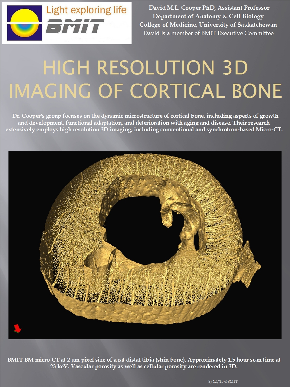
Posted on March 31, 2011
The dynamic microstructure of cortical bone Growth and development, functional adaptation, and deterioration with age and disease.
Options for Computed Tomography X-ray Imaging on BMIT
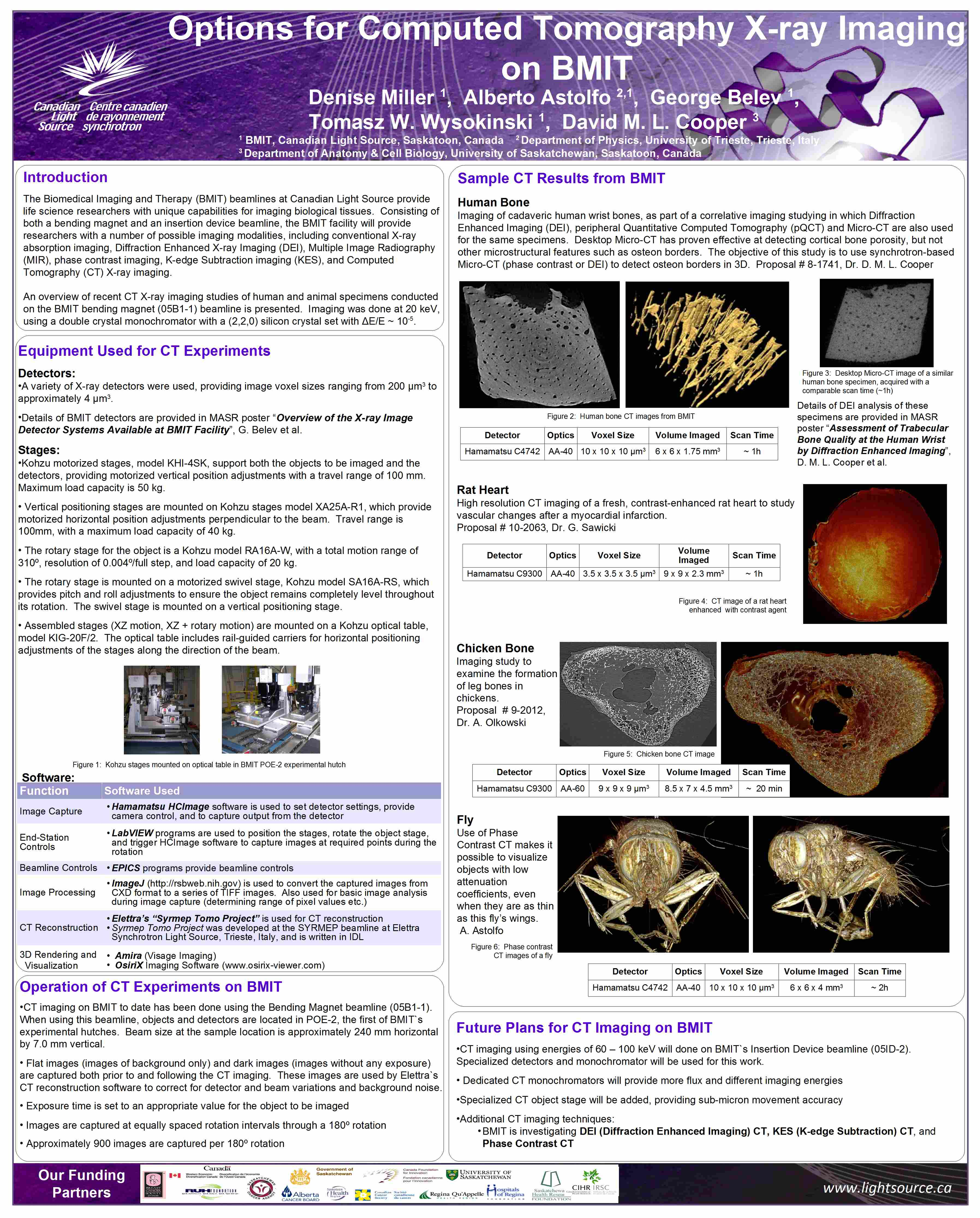
Posted on January 01, 2010
Summary of BMIT's current CT capabilities An overview of recent CT X-ray imaging studies of human and animal specimens conducted on the BMIT bending magnet (05B1-1) beamline is presented. Imaging was done at 20 keV, using a double crystal monochromator with a (2,2,0) silicon crystal set with ΔE/E ~ 10-5.
05ID-2: Superconductive Wiggler for BMIT Program
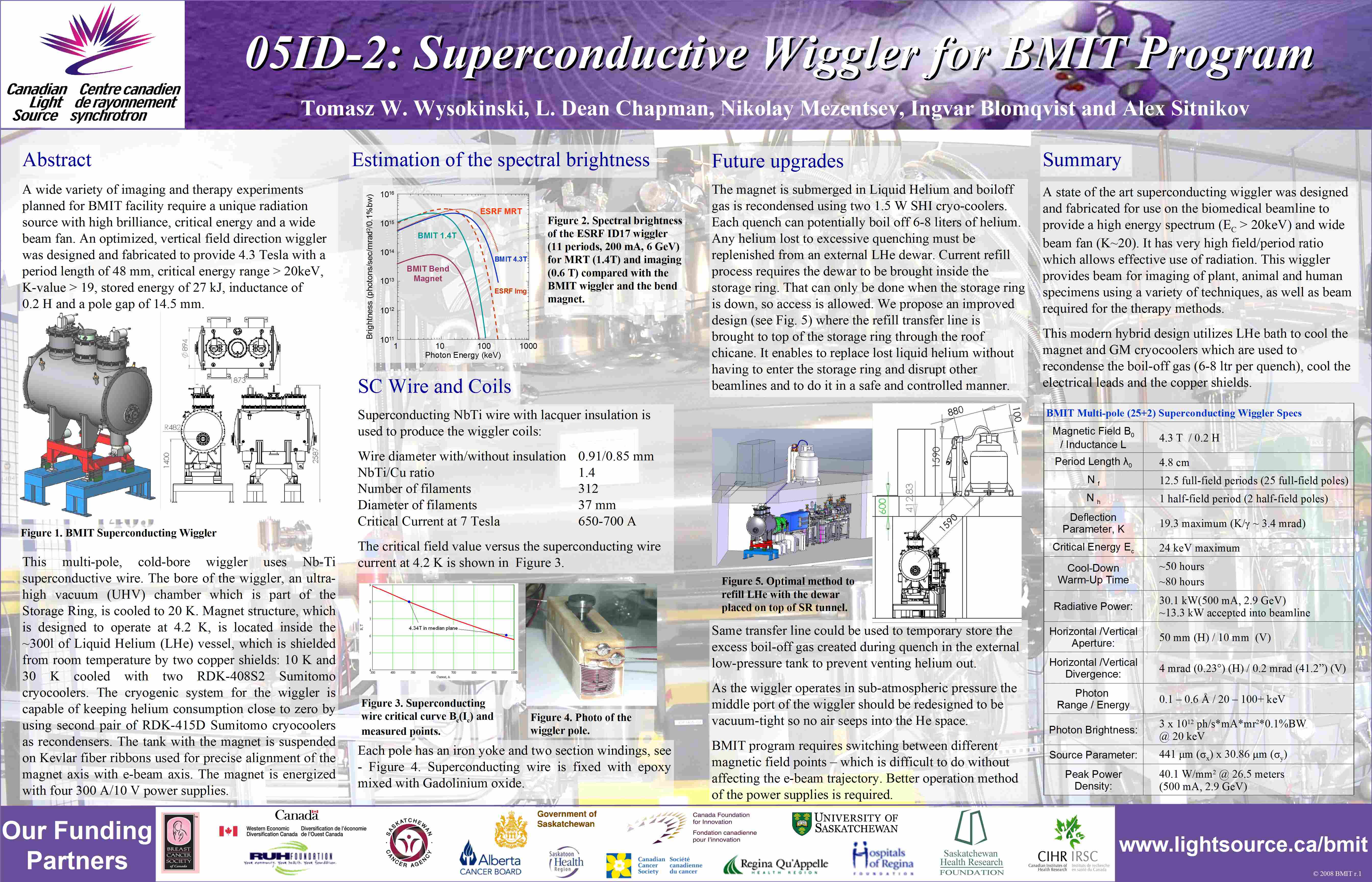
Posted on January 01, 2008
Optimized vertical field direction wiggler A wide variety of imaging and therapy experiments planned for BMIT facility require a unique radiation source with high brilliance, critical energy and a wide beam fan. An optimized, vertical field direction wiggler was designed and fabricated to provide 4.3 Tesla with a period length of 48 mm, critical energy range > 20keV, K-value > 19, stored energy of 27 kJ, inductance of 0.2 H and a pole gap of 14.5 mm.
POE-2 Gas Bremsstrahlung Studies on 05ID-2 Beamline
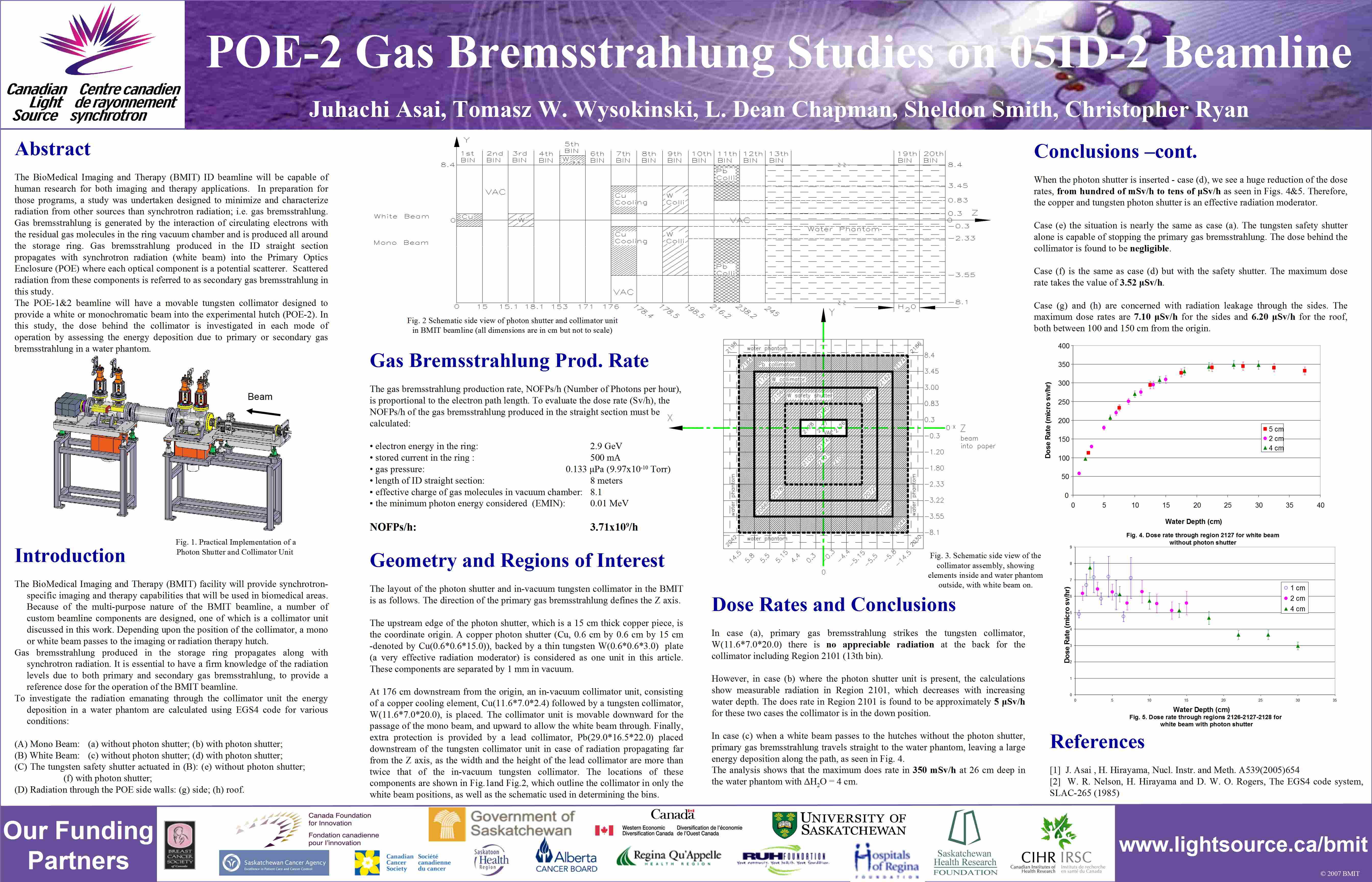
Posted on January 01, 2007
Getting rid of gas radiation The BioMedical Imaging and Therapy (BMIT) ID beamline will be capable ofhuman research for both imaging and therapy applications. In preparation forthose programs, a study was undertaken designed to minimize and characterizeradiation from other sources than synchrotron radiation; i.e. gas bremsstrahlung.Gas bremsstrahlung is generated by the interaction of circulating electrons withthe residual gas molecules in the ring vacuum chamber and is produced all aroundthe storage ring.
BMIT "MRT Lift" - A positioning system for biomedical imaging and therapy
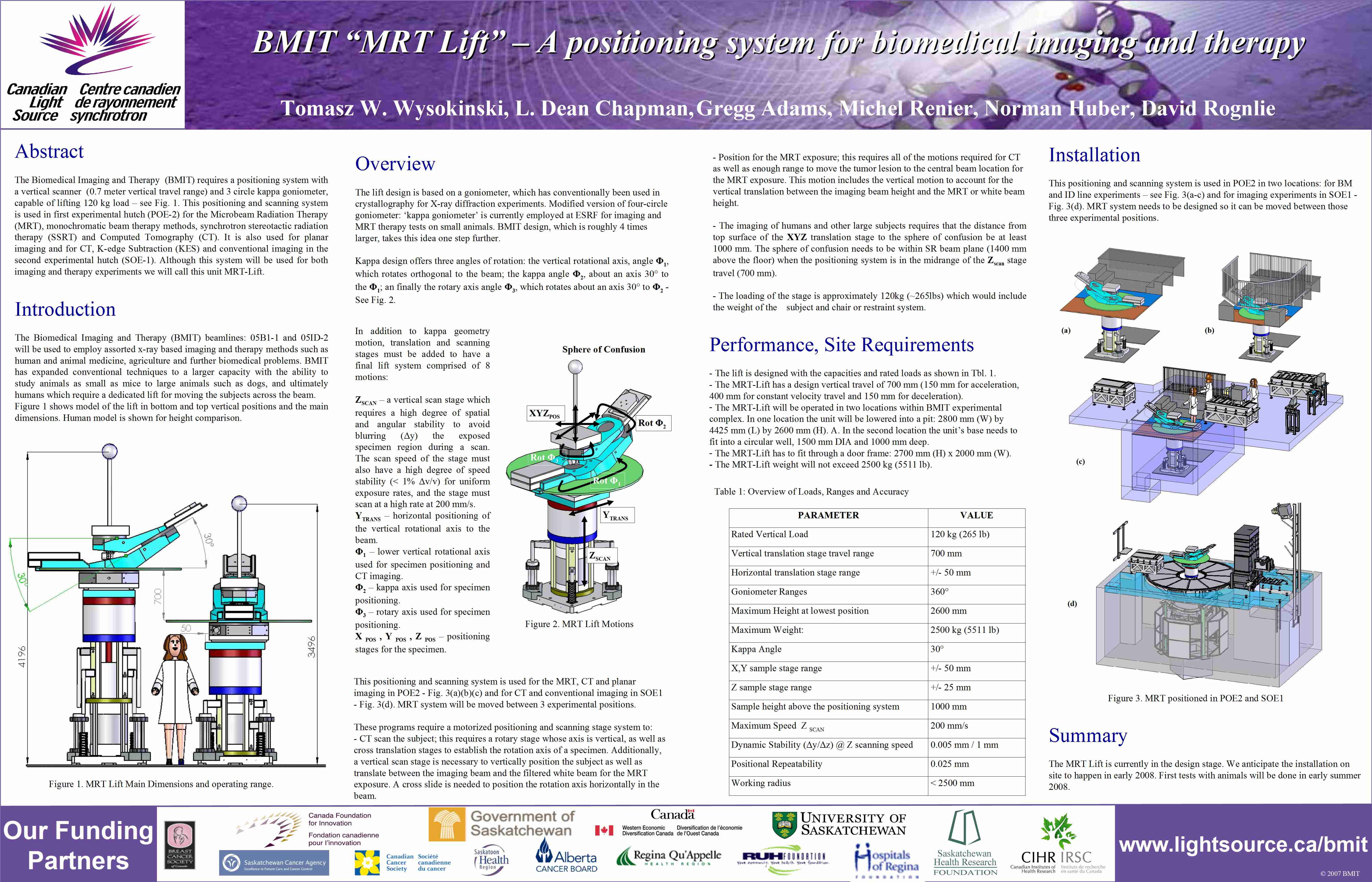
Posted on January 01, 2007
Bigger and better The MRT lift is capable of moving and lifting samples and patients larger than any previous synchrotron biomedical positioning system.
The Proposed BioMedical Imaging & Therapy Program
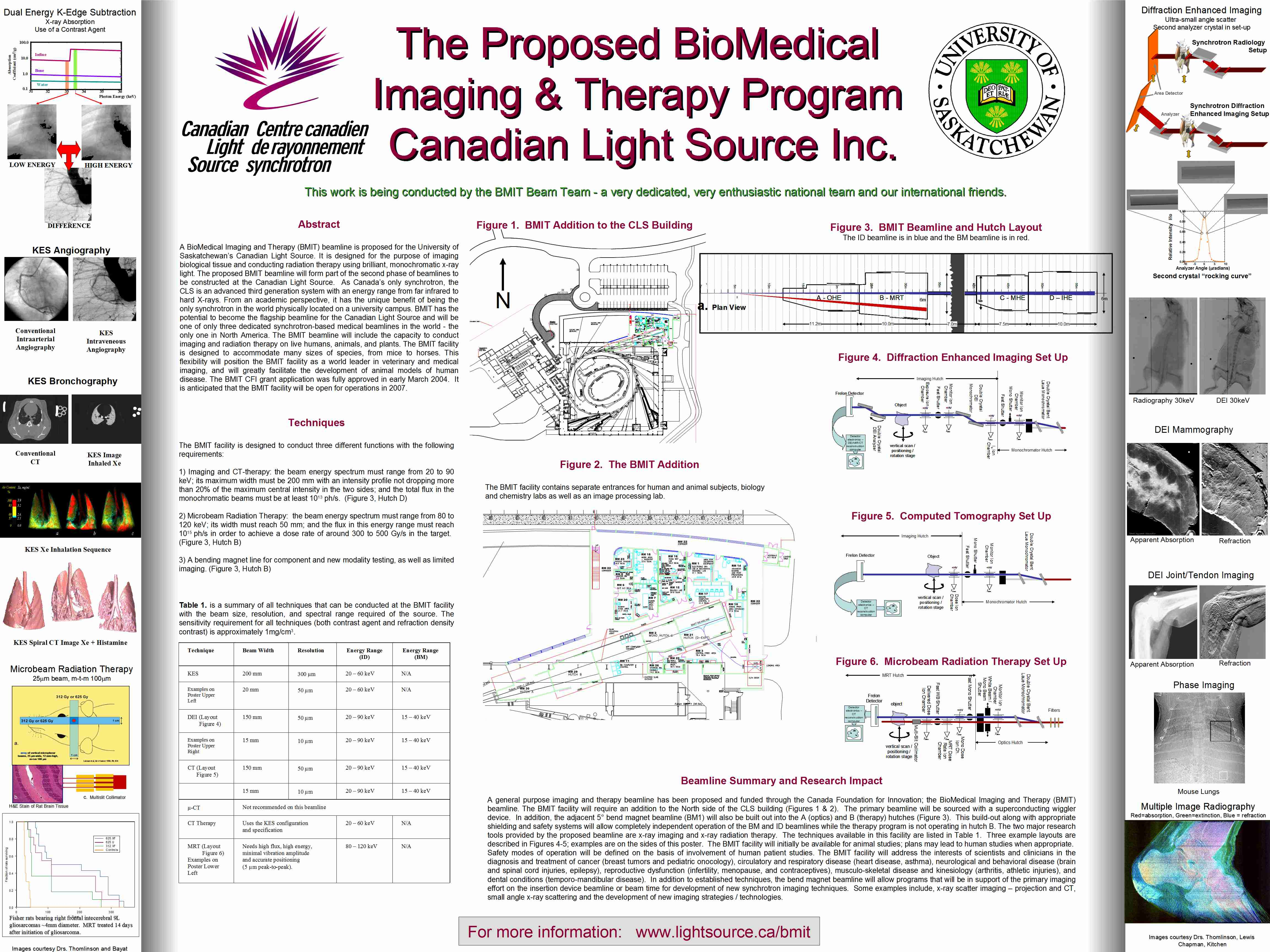
Posted on January 01, 2005
A healing light for research Designed for the purpose of imaging biological tissue and conducting radiation therapy using brilliant, monochromatic x-ray light.
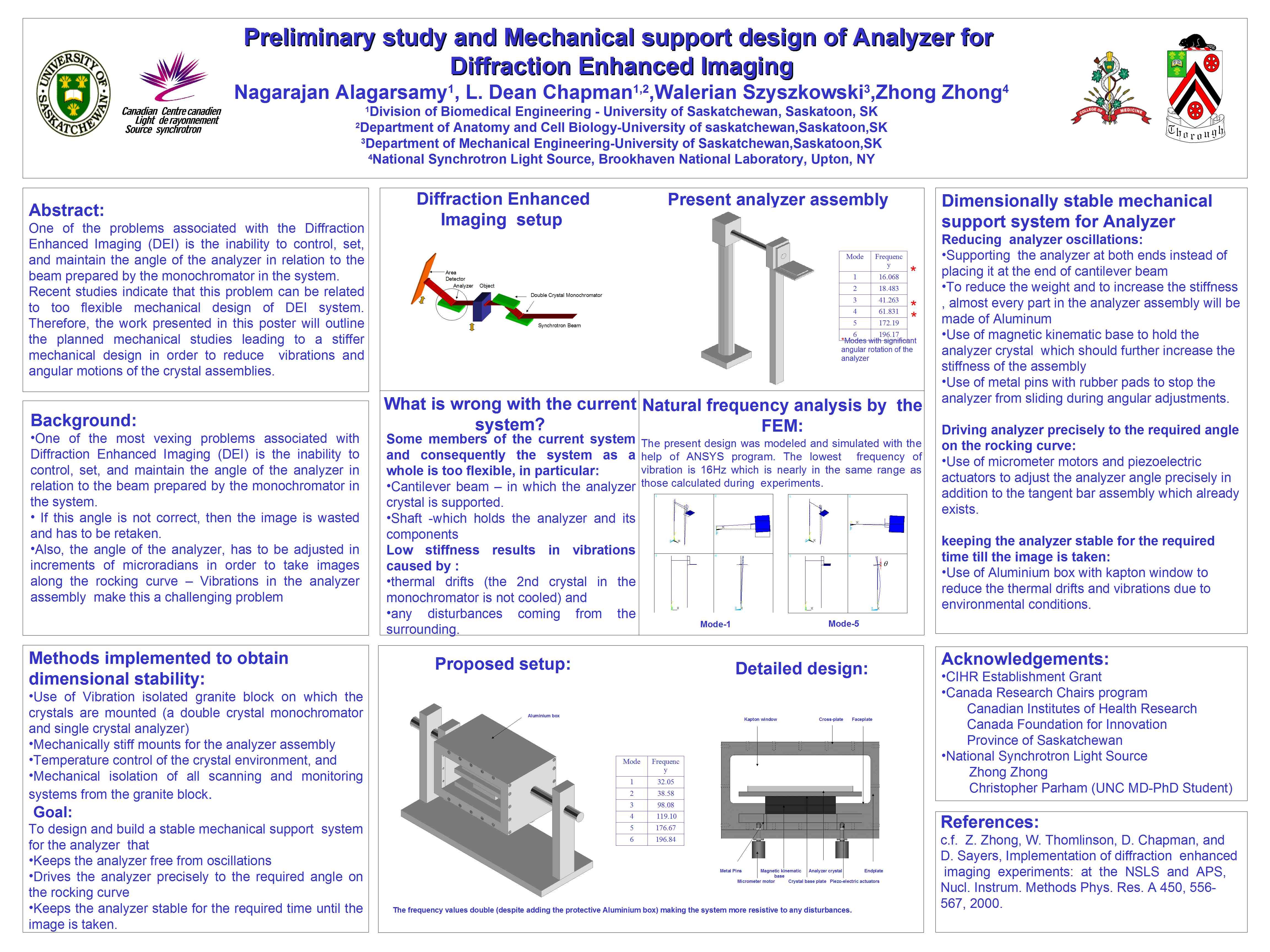
Preliminary study and Mechanical support design of Analyzer for Diffraction Enhanced Imaging
Posted on January 01, 2005
Solving problems which plague DEI Problems with the angle of the analyzer may be solved by a stiffer mechanical design.