Applications
Instruments and techniques available at BMIT have been successfully applied to numerous biomedical and preclinical studies using both excised tissue specimens and animal models ranging in size from zebra fish and mice to piglets. Several examples are shown below. For more examples please visit the research highlights page. BMIT’s powerful imaging capabilities are also routinely utilized by experts for research of alloys and composite materials, renewable energy, environmental science, and many other applications.
Scientists can utilize BMIT techniques to:
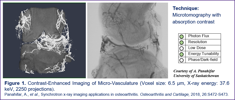
- Perform ultrafast scans. CT scans can be performed up to 100 times faster than with lab scanners (Figure 1)
- Achieve very high spatial resolution. Sample features with characteristic dimensions down to two microns can be resolved, approaching the typical resolution of histology (Figures 1, 2, 4).
- Perform low-dose temporal in-vivo imaging of animal models. The dose deposited in specimens is up to 10 times smaller than with lab scanners (Figs. 3, 6).
- Differentiate between soft tissues which do not normally exhibit contrast when imaged with conventional laboratory and clinical X-ray scanners (Figures 3, 5, 6).
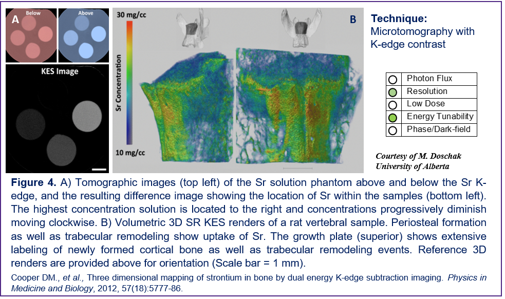
- Exploit outstanding 3D elemental sensitivity to medical contrast agents including Sr, Ag, I, Xe, Ba, Gd, Au (Figure 4). Very low concentrations of an agent can be detected.
To download a copy of the below images please click here.
Micro-Angiography in a Rat Model of Osteoarthritis
These images represent ex vivo micro-angiography, after infusion of barium/gelatine mixture, in an osteoarthritic rat 6 weeks post-surgery. Vasculature down to two voxel size in diameter can be observed.
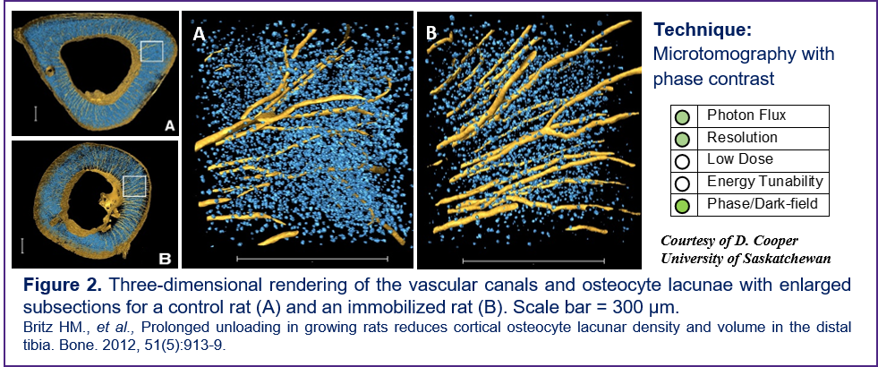
Imaging the Effects of Prolonged Unloading on Osteocyte Lacunar Density in a Rat Model
The primary objective of this study was to test the hypothesis that unloading (sciatic neurectomy) during growth results in altered osteocyte lacunar density in the tibial diaphysis of the rat. Secondarily, a potential effect of unloading on mean lacunar volume was explored.
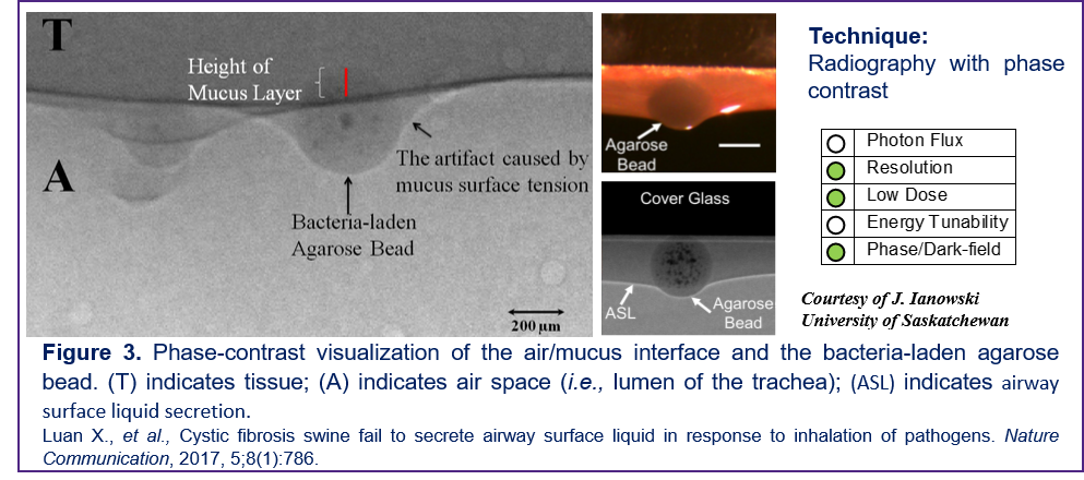
In vivo Imaging of Mucus Formation in a Piglet Model of Cystic Fibrosis
In this study the response of airway submucosal glands to inhaled bacteria and its connection to cystic fibrosis was investigated. Prior to using BMIT anatomical visualization of airway and mucus had been limited due to limitations of contrast and resolution available using conventional imaging techniques.
Mapping of Strontium Distribution in a Rat Model of Osteoporosis
K-edge subtraction (KES) imaging was used as a non-invasive imaging technique to track the 3D distribution of strontium within animal models of bone disease, and to determine the impact of strontium incorporation in mineralized bone upon mineral density and bone strength.
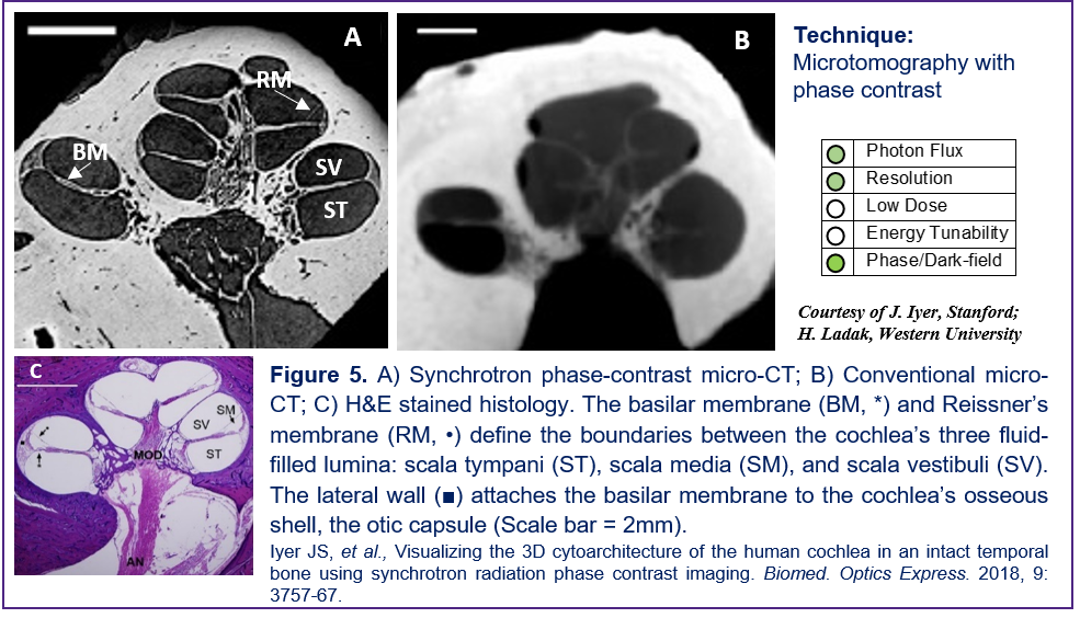
Visualizing 3D Cytoarchitecture of the Human Cochlea in an Intact Temporal Bone
This panel shows the ability of synchrotron X-ray absorption and phase contrast imaging to enable visualization of sensory cells and nerve fibers in the cochlea’s sensory epithelium in situ (3D) in intact, non-decalcified, unstained human temporal bones.
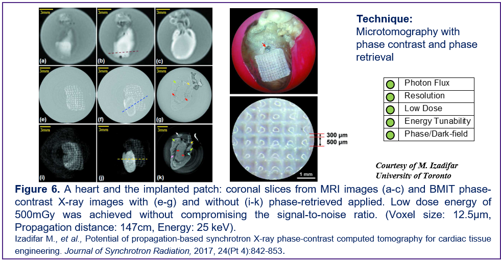
Imaging Low-Contrast Scaffolds for Cardiac Regenerative Therapy
The visualization of hydrogel-based cardiac scaffolds is essential for longitudinal studies of cardiac regenerative therapy. Synchrotron X-ray phase contrast imaging computed tomography provides significantly more information than clinical MRI (3T) to visualize the microstructural features of the hydrogel cardiac patch for scaffold-based myocardial tissue engineering.